Embryology and Anatomy of the Diaphragm
Thomas W. Shields
The diaphragm serves as the anatomic division between the thoracic and the abdominal cavities; as such, it is a muscular structure that is dealt with by both abdominal and thoracic surgeons. The surgical correction of acquired and congenital abnormalities of the diaphragm may be made from either abdominal or thoracic approaches, depending on the nature of the lesion, the location of the lesion, other abnormalities of the chest or abdomen, and the particular training and experience of the involved surgeon. The diaphragm exists as an anatomic barrier but is not a surgical barrier; a competent surgeon should be able to handle any surgical problem involving the diaphragm and therefore should be versatile enough to approach diaphragmatic lesions from either above or below.
Embryology
The diaphragm originates from an unpaired ventral portion (septum transversum), from paired dorsal lateral portions (pleuroperitoneal folds), and from an irregular medial dorsal portion (dorsal mesentery) (Fig. 51-1). The septum transversum, formed during the third week of gestation, separates the pericardial region from the rest of the body cavity. This part of the diaphragm grows dorsad from the ventral body wall and moves caudad with the other contributors to the diaphragm to reach the normal position of the diaphragm at about 8 weeks. The pleuroperitoneal folds arise on the lateral body walls, at the level where the cardinal veins swing around to enter the sinus venosus of the heart. These folds extend medially and somewhat caudad to join with the septum transversum and the dorsal mesentery to complete the development of the diaphragm at about the seventh week; the right pleuroperitoneal canal closes somewhat earlier than the left. Muscle fibers migrate from the third, fourth, and fifth cervical myotomes, carrying along their innervation, and grow between the two membranes to complete the structures of the diaphragm. During the 10th week, the intestines return from the yolk sac to the abdominal cavity and, at about 12 weeks, rotation and fixation of the intestines occur. A delay or variation in the described timetable may result in a variety of congenital hernias with or without a hernial sac or may even result in a congenital eventration of a hemidiaphragm. Early return of the intestines to the abdomen before closure of the pleuroperitoneal membrane results in a hernia through this opening (a so-called foramen of Bochdalek hernia). A sac is not usually present, but if it is, the return of the intestines may have occurred after the closure of the pleuroperitoneal membrane but before the migration of the cervical myotomes between the membranes. Foramen of Morgagni hernias occur anteriorly, almost always have a sac, and therefore probably result from lack of ingrowth of the cervical myotomes. A congenital short esophagus is related to late closure of the diaphragm and early return of the intestine to the abdomen. Congenital eventration may be a total error of ingrowth of cervical myotomes in one or both hemidiaphragms and therefore is actually a large congenital diaphragmatic hernia and not an
eventration. An absent diaphragm probably represents an error of growth of the septum transversum and other embryologic elements. Duplication of a hemidiaphragm can occur. The fusion and formation timetable variations may also involve defects in the diaphragm in association with certain vascular anomalies of the lungs and heart.
eventration. An absent diaphragm probably represents an error of growth of the septum transversum and other embryologic elements. Duplication of a hemidiaphragm can occur. The fusion and formation timetable variations may also involve defects in the diaphragm in association with certain vascular anomalies of the lungs and heart.
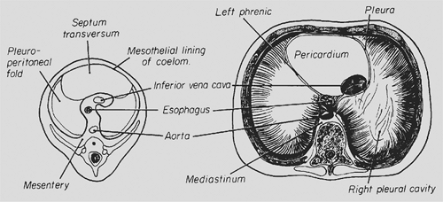 Figure 51-1. Embryologic components of the diaphragm. (Redrawn from Shields TW. The diaphragm. In Nora P, ed. Operative Surgery: Principles and Techniques. Philadelphia: Lea & Febiger, 1972.) |
Anatomy
Gross Features
The diaphragm is a dome-shaped structure of muscular fibers radiating out from either side of an irregularly shaped central tendon; it consists of the right and left hemidiaphragms. In structure and function, the diaphragm differs from any other muscle in the body. It is a muscular septum between the abdominal and thoracic cavities, serving as the major muscle of respiration. Its domelike shape allows important abdominal structures, such as the liver and the spleen, to have the protection of the lower ribs and the chest wall. The dome of the right hemidiaphragmatic leaf is normally at a higher level than is that of the left. It has been generally thought that this is the result of the liver mass beneath the right leaf. However, this view has been brought into question by the studies of Reddy and colleagues.14 Rather than the former hypothesis, their observations support the concept that the cardiac mass is responsible for the lower portion of the left hemidiaphragm.
Voluntary muscular fibers originate from the xiphisternum, from the lateral lower six ribs on each side, and from the external and internal arcuate ligaments that arise from the upper three lumbar vertebrae. Bilaterally, the muscle fibers insert into the central tendon of the diaphragm. The muscle mass of the diaphragm is considered by De Troyer and associates3 and Rochester15 as comprising two distinct parts: a thin costal muscle mass and a thicker crural portion. Although both muscle masses are innervated by the phrenic nerves, their activity on stimulation is different. The differences that result in diaphragmatic and lower chest wall movement are discussed in Chapter 52. Suffice it to mention that the movement of the crural portion has the lesser effect on ventilatory exchange.
The central tendon is a thin aponeurosis of closely interwoven fascial fibers in the form of a three-leaf clover. The two lateral leaves form the dome of the diaphragm, and the third (anterior) leaf is fused with the diaphragmatic surface of the pericardium.
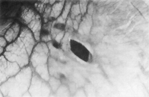 Figure 51-2. Thoracoscopy revealed a tiny hole about 3 mm in diameter in the right hemidiaphragm. Adjacent spots were made up of endometrial tissue. (Adapted from Sato M, Kase K. Catamenial pneumothorax with diaphragmatic endometriosis: a case report. J Jpn Assoc Chest Surg 1997;11:583. With permission.) |
Major interest in the muscular portion of the diaphragm centers on the two crura, which play varying roles in the formation of the esophageal hiatus. The right crus arises from the bodies of the first and second lumbar vertebrae, and the fibers divide as they pass to the left, normally overlapping in front and behind to form the entire esophageal hiatus. Collis and associates,2 however, found this arrangement in only a little more than half of their subjects. In the others, the left crus contributed to a varying degree to the makeup of the hiatus, and in about 2%, the left crus made up the major portion of the esophageal hiatus.
The hiatal opening is situated at the level of the 10th thoracic vertebra just to the left of the midline and just ventral to where the aorta passes into the abdomen. The inferior vena cava passes through the tendinous portion of the right side of the diaphragm between the anterior leaf and the right lateral leaf at the level of the eighth thoracic vertebra. The other normal openings are the parasternal foramina (i.e., the foramina of Morgagni) through which the internal mammary arteries pass into the abdomen to become the superior epigastric arteries. Evidence suggests that, in some subjects, a variable number of fenestrations or pores are present or are potentially so in either hemidiaphragmatic leaf. These are more commonly observed on the right than on the left and are located posteriorly when present. These fenestrations are of no importance normally but may become the route of fluid or air to traverse from the peritoneal cavity into the pleural space, as demonstrated by the studies of Park and Pham13 and Urhahn and Gunther22 (Fig. 51-2).
The thoracic side of the diaphragm is covered with the parietal pleura and the abdominal surface with peritoneum except at the naturally occurring openings, and the bare area is occupied by a portion of the liver.
Blood Supply
The principal blood supply of the diaphragm is derived directly from the aorta or from its most superior abdominal branches (Fig. 51-3); its venous drainage empties into the inferior vena cava. Both the arterial supply and the venous drainage (the right and left inferior phrenic veins) are found on the undersurface
of the diaphragm (Fig. 51-4). The inferior phrenic artery usually bifurcates posteriorly near the dome of the diaphragm, and the branches course along the margins of the central tendon. The smaller posterior division courses laterally above the dorsal and lumbocostal origin of the diaphragm, where it has collateral anastomoses with the lower five intercostal arteries. The larger anterior division runs anterosuperiorly to the edge of the central tendon, where it anastomoses freely with the pericardiacophrenic artery. The venous pattern is similar except that the veins generally course along the posterior aspect of the central tendon to join the inferior vena cava. Veins on the inferior surface of the diaphragm communicate with the hepatic veins through the left triangular and coronary ligaments of the liver.
of the diaphragm (Fig. 51-4). The inferior phrenic artery usually bifurcates posteriorly near the dome of the diaphragm, and the branches course along the margins of the central tendon. The smaller posterior division courses laterally above the dorsal and lumbocostal origin of the diaphragm, where it has collateral anastomoses with the lower five intercostal arteries. The larger anterior division runs anterosuperiorly to the edge of the central tendon, where it anastomoses freely with the pericardiacophrenic artery. The venous pattern is similar except that the veins generally course along the posterior aspect of the central tendon to join the inferior vena cava. Veins on the inferior surface of the diaphragm communicate with the hepatic veins through the left triangular and coronary ligaments of the liver.
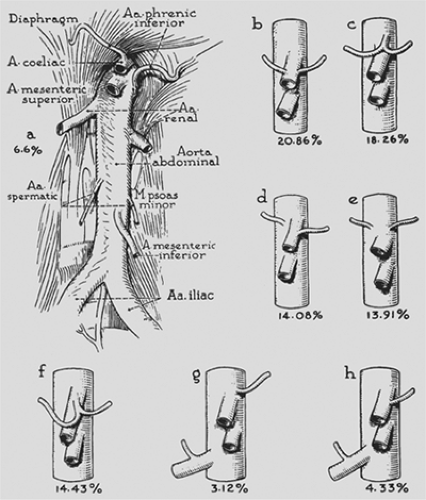 Figure 51-3. The arterial supply of the diaphragm from the abdominal aorta with variations in the origin of the inferior phrenic arteries. (Redrawn from Anson FJ, McVay C. Surgical Anatomy, 5th ed. Philadelphia: Saunders, 1971. With permission.) |
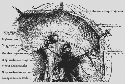 Figure 51-4. The arterial and venous distribution on the undersurface of the diaphragm. (Redrawn from Anson FJ, McVay C. Surgical Anatomy, 5th ed. Philadelphia: Saunders, 1971. With permission.) |
Nerve Distribution
The right and left phrenic nerves arise from their respective third, fourth, and fifth cervical nerve roots and constitute the total nerve supply for the ipsilateral hemidiaphragm. The distribution of each nerve is important in reference to incisions into the diaphragm. The course of each has been described by Merendino and colleagues.10 The right phrenic nerve reaches the diaphragm just lateral to the inferior vena cava and the left is just lateral to the left border of the heart. Generally the nerves divide, either just above or at the level of the diaphragm, into several terminal branches. Some are distributed to the pleural and peritoneal surfaces, but the great bulk of each nerve passes into or through the diaphragm and most often divides into four major rami to supply the various muscular portions. Usually, two of the rami share a common trunk for a varying distance so that three muscular branches arise from each phrenic nerve: one anteromedially, one laterally, and the remaining one posteriorly (Fig. 51-5). Injury to any of these branches causes paralysis of the supplied portion of the hemidiaphragm.
Stay updated, free articles. Join our Telegram channel

Full access? Get Clinical Tree


