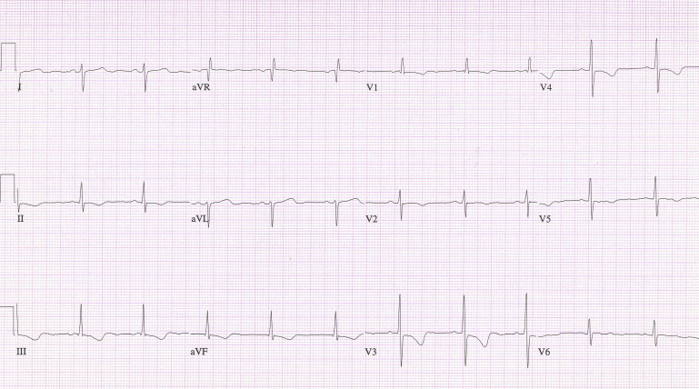
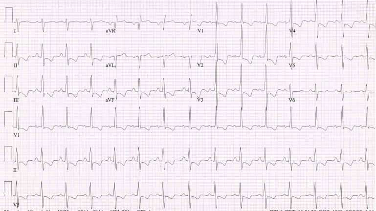
A young woman with an atrial septal defect and the Eisenmenger syndrome has worsening symptoms and electrocardiographic changes of right ventricular hypertrophy five years later.
Right-axis deviation with an rsR′s′ configuration in lead V 1 in a young woman suggests an atrial septal defect ( Figure 1 ). Fossa ovalis defect is the commonest form of atrial septal defect, but the leftward axis of the P wave (0° in the frontal plane) hints that the defect might be of the sinus venosus type. The striking signs of right ventricular hypertrophy (RVH), that is, QRS axis of +120°, dominant R′ in lead V 1 , and repolarization changes of RVH in the inferior leads as well as leads V 1 to V 5 indicate that this is not the usual secundum atrial septal defect (ASD) with normal or minimally elevated pulmonary arterial pressures. Indeed, an echo-Doppler study confirms that this patient has the Eisenmenger syndrome with increased pulmonary vascular resistance and a bidirectional shunt at the atrial level.

Eisenmenger syndrome is uncommon in patients with secundum ASDs and, especially in a young woman, must be differentiated from the also-rare primary pulmonary hypertension with a stretched foramen ovale. In contrast to the Eisenmenger syndrome with a ventricular septal defect or patent ductus arteriosus where the relative resistances in the systemic and pulmonary circulations determine the direction of flow across the communication between the 2 circulations, with an ASD the direction of flow is determined by the relative compliances of the 2 ventricles during diastole. Because of this, flow may become bidirectional or right to left when pulmonary vascular resistance is less than systemic vascular resistance.
Eisenmenger physiology virtually always grows worse. Four and a half years, later the patient was clinically worse, and the electrocardiogram signs of RVH were more striking ( Figure 2 ).


