After coronary arterial bypass operation for angina pectoris without prior myocardial infarction, a 39-year-old woman has an electrocardiogram during right ventricular pacing that is highly specific for anterior myocardial infarction.
A 39-year-old woman has a coronary arterial bypass operation for angina pectoris. She has never had a myocardial infarct. A-V sequential pacing routinely begins before the patient leaves the operating room. Figure 1 shows an early postoperative electrocardiogram.
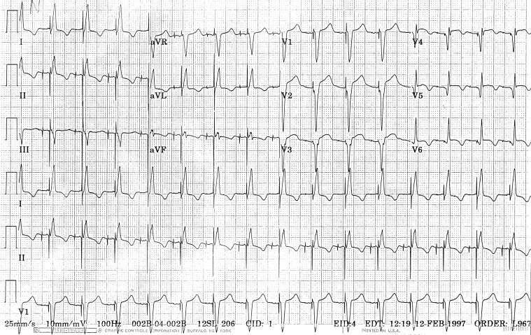
The pacemaker sequentially captures the atria and ventricles in a normal fashion at a rate of 98 beats/min. Q waves, ST-segment elevations, and/or T-wave inversions in leads I, II, aVL, and V 1 -V 6 suggest an acute anterolateral myocardial infarct not present on preoperative electrocardiograms.
The effect of the ventricular lead of an electronic pacemaker stimulating the right ventricle is a left bundle branch block–like QRS configuration, and the ST criteria for diagnosing an acute infarct are the same as in a patient with left bundle branch block: ST elevation >0.1 mV concordant with the QRS, ST depression >0.1 mV in leads V 1 , V 2 , or V 3 , or >0.5 mV ST elevation discordant with the QRS complex. Our patient has the first of these criteria with >0.1 mV of ST-segment elevation concordant with the QRS in leads V 5 and V 6 .
Because the ST-T changes in this patient could be the result of postoperative pericarditis, the QRS changes are important in making the diagnosis of myocardial infarction. As first described by Castellanos et al, when the right ventricle is being paced, the pattern of a pacemaker stimulus followed by a qR (St-qR) is quite specific for anterior wall acute myocardial infarction, especially when the St-qR pattern is seen in leads V 5 and V 6 ( Figure 1 ). Kafka found the specificity of the St-qR pattern to be 100%, but sensitivity to be low, as is the case with most “diagnostic” findings, electrocardiographic and otherwise.
Disclosures
The authors have no conflicts of interest to disclose.
See page 624 for disclosure information.
Stay updated, free articles. Join our Telegram channel

Full access? Get Clinical Tree


