Duplex Scanning and Spectral Analysis of Carotid Bifurcation Atherosclerotic Disease


Duplex Scanning and Spectral Analysis of Carotid Bifurcation Atherosclerotic Disease


Andrew N. Nicolaides, Maura Griffin and Nicos Labropoulos


In the 1980s and 1990s, grading of the degree of internal carotid artery (ICA) stenosis with duplex scanning depended on Doppler velocities at the site of maximum stenosis. With increasing resolution and image quality of gray-scale images and better color flow, plaque imaging has become complementary to velocity measurements, and in certain cases, equally important. The aim of this chapter is to present the current state of the art of duplex scanning in patients with occlusive disease at the carotid bifurcation.


Important Concepts


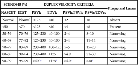
Guidelines recommend expressing the degree of ICA stenosis as a percentage of stenosis in relation to the distal normal ICA (NASCET [North American Symptomatic Carotid Endarterectomy Trial] stenosis) in routine clinical practice (see Chapter 6). Expressing the stenosis as a percentage of the bulb diameter (ECST [European Carotid Surgery Trial] stenosis) is needed in certain studies because it reflects the plaque volume and it is directly related to stroke risk. To avoid confusion, it should be remembered that the same velocities can be expressed in both NASCET and ECST grades of stenosis (Table 1).


The Doppler velocity recorded at the site of maximum stenosis depends on the pressure gradient across the stenosis. This pressure gradient is a function of both the severity of the stenosis and the collateral circulation. Let us assume that three patients have a 70% diameter angiographic stenosis of the left ICA. The right carotid bifurcation is normal in the first patient, but in the second it is occluded. In the third patient the right carotid bifurcation is normal, but the circle of Willis is incomplete. In these three patients the velocities at the stenosis in the left ICA are different because the pressure gradients are not the same. The degree of stenosis can be overestimated in the second and third patients if the physician adheres to absolute velocities. For these reasons, the use of defined stenotic strata is recommended, and duplex scanning is found to be more accurate when lesions are classified as being above or below a single level, such as 60% stenosis or 70% stenosis.


Velocity ratios were introduced following the realization that absolute velocity criteria could be inaccurate in the presence of cardiac arrhythmia, aortic valve disease, tandem plaques, recent hemispheric stroke, carotid dilatation, or aneurysm, which could result in underestimation of the degree of stenosis. Conversely, carotid coiling or kinking, arteriovenous malformations, carotid body tumors, and contralateral severe stenosis or occlusion could produce an overestimation of the stenosis.


The criteria for different velocity ratios were initially tailored to screening and selecting patients for subsequent angiography. Thus, they had a high sensitivity at the expense of specificity. The aim was to not miss a patient with a stenosis greater than 60% (NASCET). However, subsequently, with the need to make decisions to proceed to carotid endarterectomy without angiography, the criteria were tailored to have equally high sensitivity and specificity. The criteria listed in Table 1 are based on the latter option, with emphasis on obtaining the highest combined sensitivity and specificity for surgically important grades greater than 70% (ECST) as indicated by the ECST trial, 60% (NASCET) as indicated by the Asymptomatic Carotid Atherosclerosis Study (ACAS) trial, and 70% (NASCET) as indicated by the NASCET trial.


Considerable progress has been made in the quality of ultrasound imaging since the 1990s. Ultrasound has undergone great advances, and the medical community has gained further expertise in performing carotid scans and interpreting the results through the widespread use of better technology, research, and continuing medical education. In addition, various accrediting bodies have been established, such as the Intersocietal Commission for Accreditation of Vascular Laboratories, the American Institute of Ultrasound in Medicine, and the American College of Radiology, in an attempt to improve and standardize the quality of vascular examinations. In an attempt to minimize the existing variability in the performed examination, expert panels from various faculties have suggested a series of guidelines on grading carotid stenosis with ultrasound (see later).

Stay updated, free articles. Join our Telegram channel

Aug 25, 2016 | Posted by in CARDIOLOGY | Comments Off on Duplex Scanning and Spectral Analysis of Carotid Bifurcation Atherosclerotic Disease

Full access? Get Clinical Tree

Get Clinical Tree app for offline access