Diseases of the Breast

Chapter 36 Diseases of the Breast


Anatomy


The breast lies between the subdermal layer of adipose tissue and the superficial pectoral fascia (Fig. 36-1). The breast parenchyma is composed of lobes that in turn are comprised of multiple lobules. There are fibrous bands that provide structural support and insert perpendicularly into the dermis, termed the suspensory ligaments of Cooper. Between the breast and pectoralis major muscle lies the retromammary space, a thin layer of loose areolar tissue that contains lymphatics and small vessels.


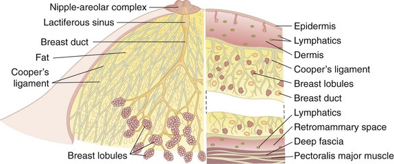
Located deep to the pectoralis major muscle, the pectoralis minor muscle is enclosed in the clavipectoral fascia, which extends laterally to fuse with the axillary fascia. The axillary lymph nodes, grouped as shown in Figure 36-2, are found within the loose areolar fat of the axilla; the number of lymph nodes is variable, depending on the size of the patient. The number of lymph nodes recovered from pathologic examination of Halsted-type radical mastectomy specimens is approximately 50 nodes.


The axillary nodes are typically described as three anatomic levels defined by their relationship to the pectoralis minor muscle. Level I nodes are located lateral to the lateral border of the pectoralis minor muscle. Level II nodes are located posterior to the pectoralis minor muscle. Level III nodes include the subclavicular nodes medial to the pectoralis minor muscle. The level III nodes are easier to visualize and remove when the pectoralis minor muscle is divided. The apex of the axilla is defined by the costoclavicular ligament (Halsted’s ligament), at which point the axillary vein passes into the thorax and becomes the subclavian vein. Lymph nodes in the space between the pectoralis major and minor muscles are termed the interpectoral group, or Rotter’s nodes, as described by Grossman and Rotter. Unless this group is specifically exposed, they are not encompassed in surgical procedures that preserve the pectoral muscles.


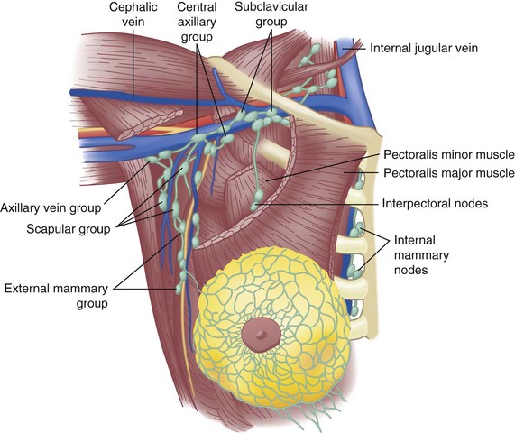
Lymphatic channels are abundant in the breast parenchyma and dermis. Specialized lymphatic channels collect under the nipple and areola and form Sappey’s plexus, named for the anatomist who described them in 1885. Lymph flows from the skin to the subareolar plexus and then into the interlobular lymphatics of the breast parenchyma. Appreciation of lymphatic flow is important for performing successful sentinel lymph node surgery (see later). Of lymphatic flow from the breast, 75% is directed into the axillary lymph nodes. A minor amount goes through the pectoralis muscle and into more medial lymph node groups, as shown in Figure 36-2. Lymphatic drainage also occurs through the internal mammary lymph nodes as the predominant drainage in up to 5% of patients and as a secondary route in combination with axillary drainage in approximately 20%. A major route of breast cancer metastasis is through lymphatic channels; the regional spread of cancer is important to understand to provide optimal locoregional control of the disease.


Coursing close to the chest wall on the medial side of the axilla is the long thoracic nerve, or the external respiratory nerve of Bell, which innervates the serratus anterior muscle. This muscle is important for fixing the scapula to the chest wall during adduction of the shoulder and extension of the arm, and division of the nerve may result in the winged scapula deformity. For this reason, the long thoracic nerve is preserved during axillary surgery. The second major nerve encountered during axillary dissection is the thoracodorsal nerve, which innervates the latissimus dorsi muscle. This nerve arises from the posterior cord of the brachial plexus and enters the axillary space under the axillary vein, close to the entrance of the long thoracic nerve. It then crosses the axilla to the medial surface of the latissimus dorsi muscle. The thoracodorsal nerve and vessels are preserved during dissection of the axillary lymph nodes. The medial pectoral nerve innervates the pectoralis major muscle and lies within a neurovascular bundle that wraps around the lateral border of the pectoralis minor muscle. The pectoral neurovascular bundle is a good landmark in that it indicates the position of the axillary vein, which is just cephalad and deep (superior and posterior) to the bundle. This neurovascular bundle should be preserved during standard axillary dissection.


The large sensory intercostal brachial or brachial cutaneous nerves span the axillary space and supply sensation to the undersurface of the upper part of the arm and skin of the chest wall along the posterior margin of the axilla. Dividing these nerves results in cutaneous anesthesia in these areas and should be explained to patients before axillary dissection. Denervation of the areas supplied by these sensory nerves can cause chronic and uncomfortable pain syndromes in a small percentage of patients. Preservation of the superiormost nerve maintains sensation to the posterior aspect of the upper part of the arm intact without compromising the axillary dissection in most patients.


Microscopic Anatomy


The mature breast is composed of three principal tissue types: (1) glandular epithelium; (2) fibrous stroma and supporting structures; and (3) adipose tissue. Lymphocytes and macrophages are also found within the breast. In adolescents, the predominant tissues are epithelium and stroma. In postmenopausal women, the glandular structures involute and are largely replaced by adipose tissue. Cooper’s ligaments provide shape and structure to the breast as they course from the overlying skin to the underlying deep fascia. Because they are anchored into the skin, infiltration of these ligaments by carcinoma commonly produces tethering which can cause dimpling or subtle deformities on the otherwise smooth surface of the breast.


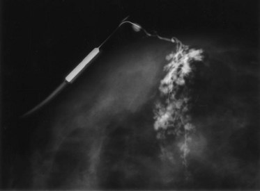
The glandular apparatus of the breast is composed of a branching system of ducts, roughly organized in a radial pattern spreading outward and downward from the nipple-areolar complex (see Fig. 36-1). It is possible to cannulate individual ducts and visualize the lactiferous ducts with contrast agents. Figure 36-3 demonstrates the arborization of branching ducts, which end in terminal lobules. The contrast dye opacifies only a single ductal system and does not enter adjacent and intertwined branches from functionally independent ductal branches. Each major duct has a dilated portion (lactiferous sinus) below the nipple-areolar complex. These ducts converge through a constricted orifice into the ampulla of the nipple.


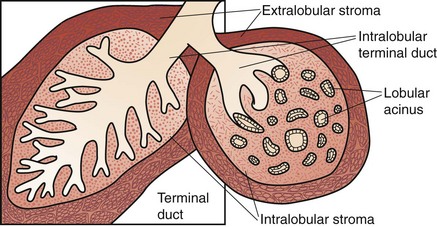
Each of the major ducts has progressive generations of branching and ultimately ends in the terminal ductules or acini (Fig. 36-4). These acini are the milk-forming glands of the lactating breast and, together with their small efferent ducts or ductules, are known as lobular units or lobules. As shown in Figure 36-4, the terminal ductules are invested in a specialized loose connective tissue that contains capillaries, lymphocytes, and other migratory mononuclear cells. This intralobular stroma is clearly distinguished from the denser and less cellular interlobular stroma and from the adipose tissue within the breast.


The entire ductal system is lined by epithelial cells, which are surrounded by specialized myoepithelial cells that have contractile properties and serve to propel milk formed in the lobules toward the nipple. Outside the epithelial and myoepithelial layers, the ducts of the breast are surrounded by a continuous basement membrane containing laminin, type IV collagen, and proteoglycans. The basement membrane layer is an important boundary in differentiating in situ from invasive breast cancer. Continuity of this layer is maintained in ductal carcinoma in situ (DCIS), also termed noninvasive breast cancer (see later, “Pathology”). Invasive breast cancer is defined by penetration of the basement membrane by malignant cells invading the stroma.


Breast Development And Physiology


Normal Development and Physiology


Prior to puberty, the breast is composed primarily of dense fibrous stroma and scattered ducts lined with epithelium. In the United States, puberty, as measured by breast development and the growth of pubic hair, begins between the ages of 9 and 12 years, and menarche (onset of menstrual cycles) begins at approximately 12 to 13 years of age. These events are initiated by low-amplitude pulses of pituitary gonadotropins, which raise serum estradiol concentrations. In the breast, this hormone-dependent maturation (thelarche) entails increased deposition of fat, the formation of new ducts by branching and elongation, and the first appearance of lobular units. This process of growth and cell division is under the control of estrogen, progesterone, adrenal hormones, pituitary hormones, and the trophic effects of insulin and thyroid hormone. There is evidence that local growth factor networks are also important. The exact timing of these events and the coordinated development of both breast buds may vary from the average in individual patients. The term prepubertal gynecomastia refers to symmetrical enlargement and projection of the breast bud in a young girl before the average age of 12 years, unaccompanied by the other changes of puberty. This process, which may be unilateral, should not be confused with neoplastic growth and is not an indication for biopsy.1


The postpubertal mature or resting breast contains fat, stroma, lactiferous ducts, and lobular units. During phases of the menstrual cycle or in response to exogenous hormones, the breast epithelium and lobular stroma undergo cyclic stimulation. It appears that the dominant process is hypertrophy and alteration of morphology rather than hyperplasia. In the late luteal (premenstrual) phase, there is an accumulation of fluid and intralobular edema. This accumulation of edema can produce pain and breast engorgement.


These physiologic changes can lead to increased nodularity and may be mistaken for a malignant tumor. Ill-defined masses in premenopausal women are generally observed through the course of the menstrual cycle prior to intervention. With pregnancy, there is diminution of the fibrous stroma and the formation of new acini or lobules, termed the adenosis of pregnancy. After birth, there is a sudden loss of placental hormones, which, combined with continued high levels of prolactin, is the principal trigger for lactation. The actual expulsion of milk is under hormonal control and is caused by contraction of the myoepithelial cells that surround the breast ducts and terminal ductules. There is no evidence for innervation of these myoepithelial cells; their contraction appears to occur in response to the pituitary-derived peptide oxytocin. Stimulation of the nipple appears to be the physiologic signal for continued pituitary secretion of prolactin and acute release of oxytocin. When breastfeeding ceases, there is a fall in the prolactin level and no stimulus for release of oxytocin. The breast then returns to a resting state and to the cyclic changes induced when menstruation resumes.


Menopause is defined by cessation in menstrual flow for at least 1 year; in the United States, it usually occurs between the ages of 40 and 55 years, with a median age of 51 years. Menopause may be accompanied by symptoms such as vasomotor disturbances (hot flashes), vaginal dryness, urinary tract infections, and cognitive impairment (possibly secondary to interruption of sleep by hot flashes). Menopause results in involution and a general decrease in the epithelial elements of the resting breast. These changes include increased fat deposition, diminished connective tissue, and the disappearance of lobular units. The persistence of lobules, hyperplasia of the ductal epithelium, and even cyst formation can all occur under the influence of exogenous ovarian hormones, usually in the form of postmenopausal hormone replacement therapy (HRT). Physicians should inquire about the menstrual history, onset of menses, and cessation of menses in postmenopausal women and record the use of HRT. HRT can lead to increased breast density, which may lower the sensitivity of mammography.


Fibrocystic Changes and Breast Pain


The condition previously referred to as fibrocystic disease represents a spectrum of clinical, mammographic, and histologic findings and is common during the fourth and fifth decades of life, generally lasting until menopause. An exaggerated response of breast stroma and epithelium to a variety of circulating and locally produced hormones and growth factors is frequently characterized by the constellation of breast pain, tenderness, and nodularity. Symptomatically, the condition is manifested as premenstrual cyclic mastalgia, with pain and tenderness to touch. This can be worrisome to many women; however, breast pain is not usually a symptom of breast cancer. Haagensen1 has recorded the symptoms of women with breast carcinoma and found pain as an unprompted symptom in 5.4% of patients. In women with breast pain and an associated palpable mass, the presence of the mass is the focus of evaluation and treatment. Normal ovarian hormonal influences on breast glandular elements frequently produce cyclic mastalgia, generally pain in phase with the menstrual cycle. Noncyclic mastalgia is more likely idiopathic and difficult to treat. Women 30 years and older with noncyclic mastalgia should undergo breast imaging with mammography in addition to a physical examination. If examination reveals a mass, this should become the focus of subsequent evaluation. Occasionally, a simple cyst may cause noncyclic breast pain, and aspiration of the cyst will usually resolve the pain. Most patients with simple cysts do not require any further evaluation unless it is a complex cyst with solid intracystic components.


Patients with fibrocystic changes have clinical breast findings that range from mild alterations in texture to dense, firm breast tissue with palpable masses. The appearance of large palpable cysts completes the picture. Mammographically, fibrocystic changes are usually seen as diffuse or focal radiologically dense tissue. By ultrasound, cysts exist in up to one third of all women 35 to 50 years of age, with most of them being nonpalpable. However, palpable cysts or multiple small cysts are typical of fibrocystic disease. Cysts, with or without fibrocystic disease, are uncommon in women older than 60 and younger than 30 years.


Histologically, in addition to macrocysts and microcysts, identified solid elements include adenosis, sclerosis, apocrine metaplasia, stromal fibrosis, and epithelial metaplasia and hyperplasia. Depending on the presence of epithelial hyperplasia, fibrocystic change is classified as nonproliferative, proliferative without atypia, or proliferative with atypia. All three alterations can occur alone or in combination and to a variable degree and, in the absence of epithelial atypia, represent the histologic spectrum of normal breast tissue. However, atypical epithelial hyperplasia (atypical ductal hyperplasia [ADH]) is a risk factor for the development of breast cancer. Atypical proliferations of ductal epithelial cells confer increased risk for breast cancer; however, fibrocystic change is not itself a risk factor for the development of breast malignancy.


Abnormal Development and Physiology


Gynecomastia


Hypertrophy of breast tissue in men is a clinical entity for which there is frequently no identifiable cause. Pubertal hypertrophy occurs in boys between the ages of 13 years and early adulthood and senescent hypertrophy is diagnosed in men older than 50 years. Gynecomastia in teenage boys is common and may be bilateral or unilateral. Unless it is unilateral or painful, it may pass unnoticed and regress with adulthood. Pubertal hypertrophy is generally treated by observation without surgery. Surgical excision may be discussed if the enlargement is unilateral, fails to regress, or is cosmetically unacceptable. Hypertrophy in older men is also relatively common. The enlargement is frequently unilateral, although the contralateral breast may enlarge with time. A number of commonly used medications, such as digoxin, thiazides, estrogens, phenothiazines, and theophylline, may exacerbate senescent gynecomastia. In addition, gynecomastia may be a systemic manifestation of hepatic cirrhosis, renal failure, and malnutrition. In both age groups, the mass is smooth, firm, and symmetrically distributed beneath the areola. It is frequently tender, which is often the reason for seeking medical attention. Both pubertal and senescent gynecomastia may be managed nonoperatively and can be fully characterized with ultrasonography. There is little confusion with carcinoma occurring in the male breast. Carcinoma is not usually tender, is asymmetrically located beneath or beside the areola, and may be fixed to the overlying dermis or to the deep fascia. A dominant mass suspected of being carcinoma should be examined with core needle biopsy. Mammography and ultrasound can also be useful tools to discriminate between gynecomastia and a suspected malignancy of the breast in older men.


Nipple Discharge


The appearance of discharge from the nipple of a nonlactating woman is a relatively common condition and is rarely associated with an underlying carcinoma. In one review of 270 subareolar biopsies for discharge coming from one identifiable duct and without an associated breast mass, carcinoma was found in only 16 patients (5.9%). In these cases, the fluid was bloody or tested strongly positive for occult hemoglobin. In another series of 249 patients, breast carcinoma was found in 10 (4%). In 8 of these patients, a mass lesion was identified in addition to the discharge. In the absence of a palpable mass or a suspicious mammogram, discharge is rarely associated with cancer.


It is important to establish whether the discharge comes from one breast or from both breasts, whether it comes from multiple duct orifices or from just one, and whether the discharge is grossly bloody or contains blood. A milky discharge from both breasts is termed galactorrhea. In the absence of lactation or a history of recent lactation, galactorrhea may be associated with increased production of prolactin. Radioimmunoassay for serum prolactin is diagnostic. However, true galactorrhea is rare and is diagnosed only when the discharge is milky (contains lactose, fat, and milk-specific proteins). Unilateral discharge coming from one duct orifice is often treated surgically when there is a significant amount of discharge (Fig. 36-5). However, the underlying cause is rarely a breast malignancy.


The most common cause of spontaneous nipple discharge from a single duct is a solitary intraductal papilloma in one of the large subareolar ducts under the nipple. Subareolar duct ectasia producing inflammation and dilation of large collecting ducts under the nipple is common and usually involves discharge from multiple ducts. Cancer is a very unusual cause of discharge in the absence of other signs. In summary, nipple discharge that is bilateral and comes from multiple ducts is not usually a surgical problem. Bloody discharge from a single duct often requires surgical excision to establish a diagnosis and control the discharge. A diagnosis of intraductal papilloma is found in most of these cases.


Galactocele


A galactocele is a milk-filled cyst that is round, well circumscribed, and easily movable within the breast. It generally occurs after the cessation of lactation or when feeding frequency has curtailed significantly. Haagensen1 has reported that galactoceles may occur up to 6 to 10 months after breastfeeding has ceased. The pathogenesis of galactocele is unknown, but it is thought that inspissated milk within ducts is responsible. The tumor is usually located in the central portion of the breast or under the nipple. Needle aspiration produces thick creamy material that may be tinged dark green or brown. Although it appears purulent, the fluid is sterile. Treatment is needle aspiration, and withdrawal of thick milky secretion confirms the diagnosis; surgery is reserved for cysts that cannot be aspirated or those that become infected.


Diagnosis Of Breast Disease


Physical Examination


The examination begins with the patient in the upright sitting position with careful visual inspection for obvious masses, asymmetries, and skin changes. The nipples are inspected and compared for the presence of retraction, nipple inversion, or excoriation of the superficial epidermis such as that seen with Paget’s disease (see Fig. 36-5A). The use of indirect lighting can unmask subtle dimpling of the skin or nipple caused by a carcinoma that places Cooper’s ligaments under tension (see Fig. 36-5B). Simple maneuvers such as stretching the arms high above the head or tensing the pectoralis muscles may accentuate asymmetries and dimpling. If carefully sought, dimpling of the skin or nipple retraction is a sensitive and specific sign of underlying cancer.


Edema of the skin produces a clinical sign known as peau d’orange (see Fig. 36-5D). When combined with tenderness, warmth and swelling of the breast, these signs and symptoms are the hallmark of inflammatory carcinoma and may be mistaken for acute mastitis. The inflammatory changes and edema are caused by obstruction of dermal lymphatic channels with emboli of carcinoma cells. Occasionally, a bulky tumor may produce obstruction of lymph channels that results in overlying skin edema. This is not typically the case with inflammatory carcinoma, where there is usually no discrete palpable mass but diffuse changes throughout the breast parenchyma. In 40 patients with inflammatory carcinoma described by Haagensen,1 erythema and edema of the skin were present in all cases, a palpable mass or localized induration was noted in 19 patients, and no localized tumor was present in 21 patients.


Involvement of the nipple and areola can occur with carcinoma of the breast, especially when the primary tumor is located in the subareolar position. Direct involvement may result in retraction of the nipple. Flattening or inversion of the nipple can be caused by fibrosis in certain benign conditions, especially subareolar duct ectasia. In these cases, the finding is frequently bilateral and the history confirms that the condition has been present for many years. Unilateral retraction or retraction that develops over a period of weeks or months is more suggestive of carcinoma. Centrally located tumors that go undetected for a long period of time may directly invade and ulcerate the skin of the areola or nipple. Peripheral tumors may distort the normal symmetry of the nipples by traction on Cooper’s ligaments.


A condition of the nipple that is commonly associated with an underlying breast cancer is Paget’s disease. First described by Sir James Paget in 1874, Paget’s disease has histologically distinct changes within the dermis of the nipple. There is often an underlying intraductal carcinoma in the large sinuses just under the nipple (see Fig. 36-5A). Carcinoma cells invade across the junction of epidermal and ductal epithelial cells and enter the epidermal layer of the skin of the nipple. Clinically, this produces a dermatitis that may appear eczematoid and moist or dry and psoriatic. It begins in the nipple, although it can spread to the skin of the areola. Many benign skin conditions such as eczema frequently begin on the areola, whereas Paget’s disease originates on the nipple and secondarily involves the areola.


Palpation of the breast tissue and regional lymph nodes follows visual inspection. While the patient is still in the sitting position, the examiner supports the patient’s arm and palpates each axilla to detect the presence of enlarged axillary lymph nodes. The supraclavicular and infraclavicular spaces are similarly palpated for enlarged nodes. Palpation of the breast is always done with the patient lying supine on a solid examining surface, with the arm stretched above the head. Palpation of the breast while the patient is sitting is often inaccurate because the overlapping breast tissue may feel like a mass or a mass may go undetected within the breast tissue. The breast is best examined with compression of the tissue toward the chest wall, with palpation of each quadrant and the tissue under the nipple-areolar complex. Palpable masses are characterized according to their size, shape, consistency, and location and whether they are fixed to the skin or underlying musculature. Benign tumors, such as fibroadenomas and cysts, can be as firm as carcinoma; usually, these benign entities are distinct, well circumscribed, and movable. Carcinoma is typically firm but less circumscribed, and moving it produces a drag of adjacent tissue. Cysts and fibrocystic changes can be tender with palpation of the breast; however, tenderness is rarely a helpful diagnostic sign. Most palpable masses are self-discovered by patients during casual or intentional self-examination.


Biopsy


Fine-Needle Aspiration Biopsy


Fine-needle aspiration (FNA) biopsy is a common tool used in the diagnosis of breast masses. It can be done with a 22-gauge needle, an appropriate-sized syringe, and an alcohol preparation pad. The aspirate must be properly prepared on a slide for cytologic examination to be clinically useful. The main usefulness of FNA biopsy is differentiation of solid from cystic masses, but it may be performed whenever a new, dominant, unexplained mass is found in the breast. The routine performance of FNA to distinguish solid from cystic breast masses has largely been replaced by ultrasonography. With a mammographically identified mass or a palpable mass, ultrasonography can quickly discriminate solid from cystic masses, which this may often obviate the need for aspiration. Cyst fluid is usually turbid and dark green or amber and can be discarded if the mass totally disappears and the fluid is not bloody. If the FNA of a suspected cyst does not reveal cyst fluid, the next step to consider is a core needle biopsy, usually with mammographic or ultrasonographic guidance. If the cyst aspiration reveals blood-tinged fluid or fluid is produced but the mass fails to resolve completely, consideration should be given to pneumocystography or image-guided core needle biopsy. It is not uncommon for cysts to reaccumulate fluid after initial aspiration. If the cyst is demonstrated to be a simple cyst on breast imaging, no further intervention is required. If the cyst is classified as a complex cyst, further imaging and evaluation should be considered to rule out an underlying carcinoma.


If the mass is solid and the clinical situation is consistent with carcinoma, cytologic examination of the aspirated material is performed. The needle is repeatedly inserted into the mass while constant negative pressure is applied to the syringe. Suction is released and the needle is withdrawn. The scanty fluid and cellular material within the needle are submitted in physiologically buffered saline (Normosol) or fixed immediately on slides in 95% ethyl alcohol. Because the cytologic evaluation will not discriminate between noninvasive and invasive breast cancers, most clinicians recommend core needle biopsy for definitive histologic diagnosis prior to surgical intervention. A positive result on FNA biopsy allows the surgeon to begin informed discussions with the patient; however, definite plans for treatment should be based on the histologic diagnosis from a core needle biopsy.


Core Needle Biopsy


Core needle biopsy is the method of choice to sample nonpalpable, image-detected breast abnormalities. This technique is also preferred for the diagnosis of palpable lesions. Core needle biopsy can be performed under mammographic (stereotactic), ultrasonographic, or magnetic resonance imaging (MRI) guidance. Mass lesions that are visualized on ultrasonography can be sampled under ultrasonographic guidance; calcifications and densities that are best seen on mammography are sampled under stereotactic guidance. During stereotactic core needle biopsy, the breast is compressed, most often with the patient lying prone on the stereotactic core biopsy table. A robotic arm and biopsy device are positioned by computed analysis of triangulated mammographic images. After local anesthetic is injected, a small skin incision is made and an 11-gauge core biopsy needle is inserted into the lesion to obtain the tissue sample with vacuum assistance. There are standards for the appropriate number of core samples to be obtained based on the type of abnormality being sampled and a clip should be placed to mark the site of the lesion, particularly for small lesions that may be difficult to find after extensive sampling. The specimens should be placed in a Petri dish and imaged to confirm that the targeted lesion has been adequately sampled. A similar approach is used for ultrasonographic and MRI-guided biopsy of lesions.


Specimen radiography of excised cores is performed to confirm that the targeted lesion has been sampled and to direct pathologic assessment of the tissue. A postbiopsy mammogram confirms that a defect has been created within the target lesion and that the marking clip is in the correct position. Wire localization and surgical excision are required if the lesion cannot be adequately sampled by core biopsy approaches, or if there is discordance between the imaging abnormality and pathologic findings.


Interpretation of Core Needle Biopsy Results


The limited sample size obtained by core biopsy techniques requires proper interpretation of the pathology results. Most patients undergoing core biopsy will have benign findings and may return to routine screening, with no other intervention required. If a malignancy is detected, histologic subtype, grade, and receptor status should be determined from the core biopsy sample. The patient may proceed to definitive treatment of the cancer if it is an early-stage breast cancer. Patients with locally advanced or inflammatory breast cancer should be treated with systemic chemotherapy prior to surgical intervention. Depending on the size of the imaging abnormality, approximately 10% to 20% of patients with a diagnosis of DCIS on core biopsy will be found to have some invasive carcinoma at definitive surgery.


The diagnosis of breast lesions using a minimally invasive procedure, such as core needle biopsy, is the preferred approach. The use of excisional breast biopsy as a diagnostic procedure increases costs and results in delays to definitive surgery for patients with cancer.2 Fewer than 10% of patients who undergo core biopsy will have inconclusive results and require wire-localized surgical biopsy for definitive diagnosis. Biopsy results that are not concordant with the targeted lesion (e.g., a spiculated mass on imaging and normal breast tissue on core biopsy) also require surgical excision. When ADH is found on core biopsy, surgical excision will reveal DCIS or invasive carcinoma in up to 20% of cases because of difficulty distinguishing ADH and DCIS in a limited tissue sample. A finding of a cellular fibroadenoma on core biopsy requires excision to rule out a phyllodes tumor.


Breast Imaging


Breast imaging techniques are used to detect small, nonpalpable breast abnormalities, evaluate clinical findings, and guide diagnostic procedures. Mammography is the primary imaging modality for screening asymptomatic women. During mammography, the breast is compressed between Plexiglas plates to reduce the thickness of the tissue through which the radiation must pass, separate adjacent structures, and improve resolution. Two views of each breast are obtained on a screening mammogram, mediolateral oblique and craniocaudal. A diagnostic mammogram is indicated for further evaluation of abnormalities identified on a screening mammogram or of clinical findings or symptoms. Magnification views are obtained to evaluate calcifications and compression views are used to provide additional detail when a mass lesion is suspected. Mammographic sensitivity is limited by breast density, with as many as 10% to 15% of clinically evident breast cancers having no associated mammographic abnormality. Digital mammography acquires digital images and stores them electronically, thereby allowing manipulation and enhancement of images to facilitate interpretation. Digital mammography appears to be superior to traditional film-screen mammography for detecting cancer in younger women and those with dense breasts. Mammography in women younger than 30 years, whose breast tissue is dense with stroma and epithelium, may produce an image without much definition. As women age, the breast tissue involutes and is replaced by fatty tissue. On mammography, fat absorbs relatively little radiation and provides a contrasting background that favors detection of small lesions. Computer-assisted diagnosis (CAD) has been shown to increase the sensitivity and specificity of mammography and ultrasound over review by the radiologist alone.


Screening Mammography


Screening mammography is performed in asymptomatic women with the goal of detecting breast cancer that is not yet clinically evident. This approach assumes that breast cancers identified through screening will be smaller, have a better prognosis, and require less aggressive treatment than cancers identified by palpation. The potential benefits of screening are weighed against the cost of screening and the number of false-positive studies that prompt additional workup, biopsies, and patient anxiety.


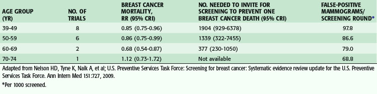
Eight prospective randomized trials of screening mammography have been performed, with almost 500,000 women participating. The impact of mammographic screening on breast cancer mortality has been assessed by age group at specific intervals by the U.S. Preventive Services Task Force and the most recent report resulted in a change in recommendations for breast cancer screening. Based on the review of eight trials in women aged 39 to 49 years, screening mammography reduced the risk for breast cancer death by 15% (relative risk [RR], 0.85; credible interval [CrI], 0.75 to 0.96). In the six trials that included women aged 50 to 59, there was a reduction in risk of 14% (RR, 0.86; CrI, 0.75 to 0.99). There were two trials that included women aged 60 to 69 and, in this age group, there was a reduction in risk of 32% (RR, 0.68; CrI, 0.54 to 0.87). There was only one trial that included women older than 70 years, and it was concluded that there were insufficient data to recommend routine screening in this age group. Based on these results, the most recent U.S. Preventive Services Task Force report recommended biennial screening mammography for women aged 50 to 74 years and recommended against screening for those aged 40 to 49 and women older than 75 years of age.2 The recommendations were based on the risk reduction, number of women needed to invite for screening to prevent one breast cancer death, and potential for harm from additional testing and biopsies (Table 36-1).


< div class='tao-gold-member'>

Stay updated, free articles. Join our Telegram channel

Aug 1, 2016 | Posted by in CARDIAC SURGERY | Comments Off on Diseases of the Breast

Full access? Get Clinical Tree

Get Clinical Tree app for offline access