Diabetic Foot Ulcer
JASON CROWNER and WILLIAM MARSTON
Presentation
A 26-year-old female with past medical history of end-stage renal disease requiring dialysis, hypertension, and type 2 diabetes presents to the wound care clinic for evaluation of a left great toe ulcer. Vital signs are grossly normal except for hypertension with systolic blood pressures in the 180s. Physical examination reveals an obese African American female with palpable left foot pedal pulses and an ulcer on the plantar aspect of the great toe (Fig. 1). A large callus surrounds the ulceration with necrotic tissue present within the base and apparent drainage. The patient states the wound has been present for several months and has been treated with Polysporin and oral antibiotics without significant improvement. She also reports a history of poorly controlled diabetes with a hemoglobin A1c of 9.9, persistent tobacco use, and continued ambulation on the foot.
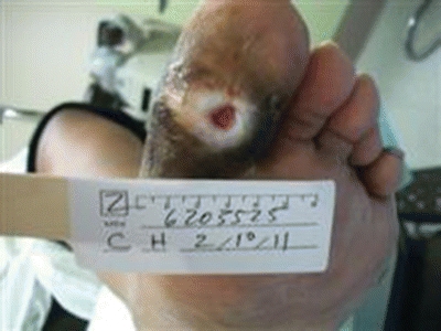
FIGURE 1 Picture showing plantar base of great toe ulcer.
Differential Diagnosis
Approximately 15% of diabetic patients will develop a full-thickness lower extremity ulcer, usually due to the combination of sensory neuropathy and motor neuropathy leading to toe and foot deformities. This is termed diabetic foot ulcer (DFU). In some cases, the condition is exacerbated by diabetes-induced peripheral arterial disease (PAD). Initial evaluation should consider the patient’s diabetes and its complications to be the main causation of the ulcer until proven otherwise. Other common causes of lower extremity ulceration include nondiabetic arterial insufficiency, chronic venous insufficiency, trauma, and chronic pressure. Unusual causes include malignancy, lupus, Raynaud’s syndrome, sickle cell disease, and gout.
In the evaluation of ulcerations present on the lower extremities, it is important to obtain a full and adequate history and physical examination. Diabetic ulcers frequently occur due to poor foot care in the setting of neuropathy, but the combined presence of PAD can exacerbate small wounds. Physical exam should include monofilament foot testing for neuropathy and noninvasive examination of the blood supply with an arterial duplex of the lower extremity. Routine laboratory evaluation should include hemoglobin A1c and plain x-rays of the foot to evaluate for osseous abnormalities or osteomyelitis.
Workup
Our patient’s radiologic evaluation of the foot demonstrated soft tissue swelling and irregularity along the great toe distal phalanx consistent with possible infectious osteitis (Fig. 2). Laboratory values demonstrated an elevation of her hemoglobin A1c of 9.9, a white blood cell count within normal limits, and wound cultures demonstrating Staphylococcus. Arterial duplex of the right lower extremity demonstrated no evidence of PAD by presence of palpable pedal pulses.
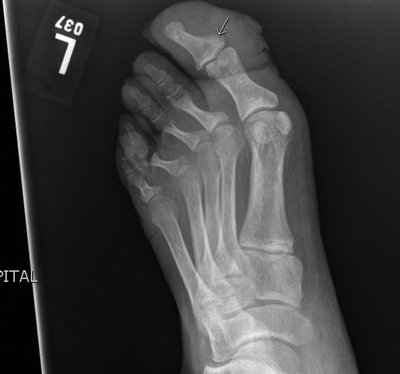
FIGURE 2 Left foot x-ray—soft tissue swelling and irregularity along the great toe consistent with possible infectious osteitis at the base of the great toe phalanx.
Discussion
Diabetic foot ulceration is defined as a full-thickness wound penetrating through the dermis (the deep vascular and collagenous inner layer of the skin) located below the ankle in a diabetic patient according to the International Working Group on the Diabetic Foot. Multiple factors play into a DFU formation and inability to heal. The major factors in the formation of DFU are secondary to peripheral neuropathy and ischemia from PAD.
The DFU can thus be categorized into purely neuropathic, purely ischemic, or a combination of the two. A combination of the two, neuroischemia, makes up the majority, with a prevalence of approximately 50%, followed by neuropathic at 35%, and ischemic at 15%. The incidence of limb loss is highest in the neuroischemic group, estimated at 15% at 5 years. Early identification and treatment of PAD has been found to reduce the risk of limb loss.
Diagnosis and Treatment
A detailed evaluation of the wound is important to document the appearance, size, location, and the state of infection. This information should be available at follow-up visits to determine whether the wound is responding to the treatment. DFUs treated with a comprehensive management plan should demonstrate a significant reduction in size within 3 to 4 weeks of initiating treatment. If not, a full reevaluation of the wound and its potential underlying causes should be performed. The examination should also include an investigation as to the specific cause of the ulceration: poor foot care, ill-fitting footwear, or bony changes causing pressure.
Operative management is warranted when there is nonviable tissue, infection, or need for improvement of blood flow. The need for wound cultures is controversial in the treatment of DFUs. In clearly infected wounds, tissue samples for culture are useful to identify the specific bacterial pathogens and guide antibiotic therapy. In wounds that do not appear to be clinically infected, it is unclear whether wound cultures are useful to identify and treat overgrowth of bacteria.
Several measures have been found to be useful in determining whether a lower extremity has adequate blood supply to heal with good wound care. Ankle-brachial indices (ABIs) are useful if the tibial blood vessels are not too calcified for compression. An ABI greater than 0.8 usually suggests that sufficient supply is available to support wound healing. An ABI between 0.5 and 0.8 is marginal, and an ABI below 0.5 is typically inadequate for an ankle or foot ulcer to heal. Patients with heavily calcified tibial vessels may yield falsely elevated ABIs. Toe pressures can be obtained with the use of a plethysmography cuff and are useful in situations where foot disease is present or ABIs are falsely elevated. An alternative to consider is to obtain transcutaneous oxygen measurement (TCOM) in the foot. A toe pressure greater than 50 mm Hg or a TCOM above 40 torr is typically associated with a favorable potential to heal, while lower numbers suggest that revascularization is likely to be necessary.
In regard to our case presentation, the patient’s physical evaluation demonstrated the presence of infection and nonviable tissue. Arterial duplex examination of the limb resulted in an ABI of 0.95 and toe pressure of 140 consistent with normal arterial supply. The decision was made for operative debridement.
Ulcer Management
Nonsurgical
Conservative management involves aggressive wound care utilizing pressure offloading, topical wound agents, dressings, and adjuvant therapies. In a patient with adequate arterial supply, the primary underlying etiology relates to neuropathy-induced pressure. Thus, pressure offloading is critical to achieving reliable wound healing. A variety of options must be employed in individual patient cases to effectively relieve pressure including custom-molded orthotics, custom-designed shoes, full-length boots, and total contact casts (Fig. 3). The participation of a skilled podiatrist and/or orthotist is important to achieve optimal healing outcomes. Once healing of the wound has occurred, it is still important to counsel the patient on the importance of proper fitting footwear and daily monitoring.
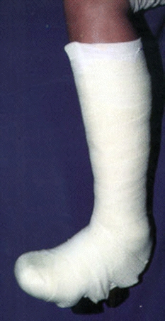
FIGURE 3 Total contact cast.


