Congenital Heart Disease in Adults


Congenital Heart Disease in Adults


Timothy B. Cotts

Julie A. Kovach

Albert P. Rocchini

Edward L. Bove


More than 1 million adults in the United States currently have congenital heart disease. Many have undergone surgery for repair or palliation of their heart defects and are cared for in their communities. Because of advances in surgical and medical care over the past several decades, an additional 9,000 patients per year with congenital heart disease reach adulthood (1). The profile of adults with congenital heart disease continues to evolve. Patients with previously nonsurvivable lesions, particularly complex single-ventricle anatomy such as hypoplastic left heart syndrome, are now reaching adulthood. This chapter reviews the clinical characteristics of the most common cardiac lesions with expected survival to adulthood: atrial septal defect, ventricular septal defect, coarctation of the aorta, patent ductus arteriosus, tetralogy of Fallot, D-transposition of the great arteries, and single-ventricle lesions palliated to the Fontan procedure.


ATRIAL SEPTAL DEFECT

Atrial septal defects (ASDs) are the most common of the congenital cardiac defects first diagnosed in adulthood, totaling approximately 30% of cases of congenital heart disease diagnosed in adults. Ostium secundum ASDs, which are defects in the region of the fossa ovalis, are most frequent and represent 75% of all ASDs. The ostium primum defect, which is absence of the lower part of the interatrial septum at the crux, occurs in 15% of patients, is typically associated with a cleft mitral valve, and is most often diagnosed in childhood. Sinus venosus defects, 10% of all ASDs, usually occur in the interatrial septum just inferior to the orifice of the superior vena cava and are associated with anomalous drainage of a pulmonary vein or veins from the right lung. In rare cases, the sinus venosus defect is of the inferior vena cava type visible below the fossa ovalis and immediately superior to the orifice of the inferior vena cava. Even more rare is the coronary sinus defect, which results from absence of part of the roof of the coronary sinus with left-to-right shunting (Fig. 36.1). Infrequently, ASD may present as part of a genetic disorder, such as Holt-Oram syndrome, also known as “heart-hand” syndrome, which is characterized by ASD, usually secundum in type, and distinctive abnormalities of the bones of the forearms, hands, and upper limbs. In regions of the world with high prevalence of rheumatic fever, affected patients may have both secundum ASD and rheumatic mitral stenosis, the combination of which is the Lutembacher complex. In these individuals, the severity of the mitral stenosis may be underestimated because of the coexisting left-to-right shunt.

A patent foramen ovale is defined as a persistent flaplike opening between the atrial septum primum and secundum at the region of the fossa ovalis, caused by incomplete sealing off after birth. Patent foramen ovale is present in up to 25% of adults, is not due to embryologic absence of septal tissue, and should not be confused with the ASD. The patent foramen ovale may become
significant if paradoxic embolization occurs through the opening.
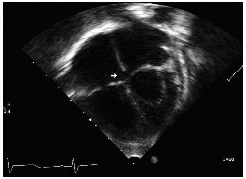


FIGURE 36.1. Apex down apical four-chamber view demonstrates a small primum atrial septal defect (arrow).


Presenting Symptoms and Signs

The physiologic hallmark of ASD is shunting of blood from the left atrium to the right atrium during late systole and early diastole. If significant, shunting produces volume overload of the right ventricle in diastole and increased flow through the pulmonary vascular bed. The magnitude of left-to-right shunting depends not only on the size of the defect but also on the relative stiffness of the left versus the right ventricles and the proportional resistances of the pulmonary and the systemic vasculatures. In an aging adult with decreased ventricular compliance due to systemic hypertension or increased left ventricular diastolic pressure from cardiomyopathy or ischemia, a previously small left-to-right shunt may increase in size and cause new symptoms. In the patient with a very large ASD, fixed pulmonary hypertension (Eisenmenger physiology) may supervene, and the shunt may reverse, becoming bidirectional or predominantly right to left. Most patients with ASD are asymptomatic until the third or fourth decade of life, unless the defect is quite large. Dyspnea on exertion manifests in 30% of patients by the third decade and in 75% by the fifth decade of life. Effort intolerance and fatigue are common. By 40 years of age, the patient frequently experiences palpitations from atrial fibrillation or flutter; less frequently, sick sinus syndrome may be present in patients with sinus venosus defects. Symptoms of systemic venous congestion and right-sided heart failure (e.g., edema, ascites) may occur in 10% of affected 40-year-olds. The patient with ostium primum ASD may initially have fatigue due to bradycardia from complete atrioventricular block. Symptoms of pulmonary congestion from uncorrected mitral regurgitation may be prominent in these patients. In rare cases, the adult with a small ASD may suffer a cerebrovascular event or peripheral emboli as a result of paradoxic embolization through the defect. Finally, the adult with Eisenmenger syndrome may be cyanotic at rest or develop cyanosis with exertion.

In the patient with a large left-to-right shunt, the jugular veins may be distended with “left atrialization” of the pressure waveform (a wave = V wave). A dynamic right ventricular impulse may be palpated at the left sternal border, and a prominent pulmonary artery pulsation may be noted at the upper left sternal margin. The first heart sound is normal to increased in intensity. The second heart sound is, as a rule, widely split, with loss of respiratory variation. Respiratory changes in systemic venous return to the right atrium are offset by reciprocal changes in the volume of the left-to-right shunt, thus ameliorating the respiratory changes in stroke volumes
of the right and left ventricles that cause physiologic splitting (2). If pulmonary hypertension is present, the pulmonic component of S2 may be loud and even palpable in the second left intercostal space. Because of increased pulmonary flow, a pulmonic systolic ejection murmur is present and often incorrectly assumed to represent an “innocent” murmur. Although common in children, a tricuspid diastolic flow rumble is rarely appreciated in adults. No murmur is produced by flow directly across the ASD because the interatrial pressure difference is small, and velocity of flow is low (Table 36.1).


Helpful Tests

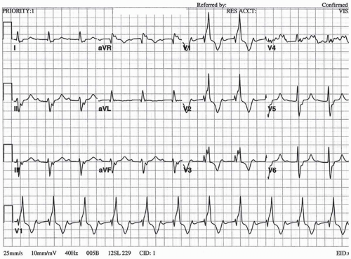
On electrocardiogram (ECG), most affected adults display normal sinus rhythm. In one study of adults with a first diagnosis of ASD, atrial fibrillation or flutter was found in 8%, complete heart block (presumably in patients with ostium primum ASD) in 1%, and complete right bundle-branch block in 8% (3). A junctional or low atrial rhythm with inverted P waves in the inferior leads may be present in patients with sinus venosus ASD that is caused by disruption of the sinus node. Classically, the ECG shows an incomplete right bundle-branch block with right axis deviation in secundum ASD and left axis deviation in primum ASD. Patients with Eisenmenger physiology may manifest right atrial or right ventricular hypertrophy (or both) with widening of the QRS interval (Fig. 36.2). On chest radiograph, more than 80% of affected adults older than 40 years display moderate to severe cardiomegaly, especially of the right atrium and ventricle. The proximal pulmonary arteries may be prominent. More than 30% of affected patients demonstrate “shunt vascularity,” with prominent small pulmonary arteries in the periphery of the lung fields (Fig. 36.3). The peripheral pulmonary arteries may be diminutive with “pruning” if fixed pulmonary vascular obstruction exists (4).

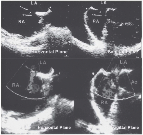
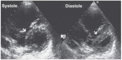
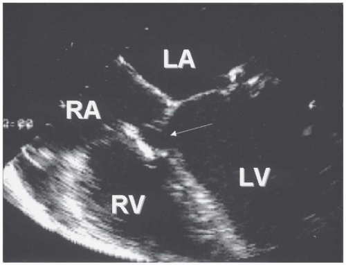
Echocardiography is the most practical and cost-effective technique for the demonstration and characterization of the anatomy and physiologic consequences of most types of ASD. Transthoracic echocardiography may demonstrate right atrial and ventricular enlargement and paradoxic septal motion consistent with volume overload of the right ventricle, and it is helpful for assessing right and left ventricular function (Fig. 36.4). The ostium primum ASD is particularly well seen on transthoracic echocardiography. “Echo dropout” of the midportion of the interatrial septum on two-dimensional echocardiography characterizes the secundum ASD, but this finding may represent a false-positive sign that is due to the relative “thinness” of the fossa ovalis in comparison to the surrounding septal tissue, especially in the four-chamber view. Color and spectral Doppler echocardiography may demonstrate flow across the defect, especially in the subcostal view, in which ASD flow is parallel to the ultrasound beam. In some patients, the secundum ASD may be visualized only after intravenous injection of agitated saline microbubbles, which are seen passing from the right atrium into the left atrium. Conversely, a “negative contrast effect,” or clear space in the bubble-filled right atrium, may be seen when unopacified blood shunts from left to right across the defect. Transesophageal echocardiography more clearly reveals the defect at the fossa ovalis in secundum defect and is superior to transthoracic echocardiography for the diagnosis of sinus venosus defects in adults (Fig. 36.5) (5,6). In addition, Doppler echocardiography demonstrates the direction of shunting, allows quantitation of the left-to-right shunt, and permits estimation of the right ventricular systolic pressure. Phase-contrast cine magnetic resonance imaging and cine computed tomography can accurately depict the morphologic features of all types of ASD and are not as invasive as transesophageal echocardiography, but they are more expensive and are not available in most practice settings (7).

Cardiac catheterization is recommended in most adults older than 40 years for characterization of coronary artery anatomy, and it is required in younger adults if fixed pulmonary vascular obstruction is suspected. In these patients, reversibility of pulmonary vascular disease should be evaluated at the time of catheterization with the use of appropriate pulmonary vasodilators, because this may influence the decision for repair.


TABLE 36.1. Clinical symptoms, signs, and evaluation of the adult with congenital heart disease


COMMON PRESENTING
SYMPTOMS


CHARACTERISTIC PHYSICAL
EXAMINATION FINDING


ECG FINDINGS


CHEST RADIOGRAPH
FINDINGS


OTHER HELPFUL TESTS


ASD


Dyspnea
Fatigue
Atrial fibrillation


Fixed split S2


Secundum ASD: IRBBB with RAD
Primum ASD: IRBBB with LAD


Cardiomegaly with shunt vascularity


Echocardiography, MRI, cardiac catheterization


VSD


Asymptomatic murmur
Cyanosis if Eisenmenger syndrome present


Systolic regurgitant murmur


RAD, RVH if Eisenmenger syndrome present


Cardiomegaly with shunt vascularity


Echocardiography, cardiac catheterization


Coarctation of the aorta


Hypertension


Blood pressure differential between upper and lower extremities


LVH


“3” sign
Rib notching


MRI or spiral CT


PDA


Asymptomatic murmur


Continuous murmur
Differential cyanosis if Eisenmenger syndrome


Normal RVH if Eisenmenger syndrome present


Normal


Echocardiography


TOF


Uncorrected: dyspnea and cyanosis


Uncorrected: single P2 pulmonic flow murmur


Uncorrected: RVH


Uncorrected: RVH with decreased pulmonary vessels


Echocardiography, MRI, cardiac catheterization


Corrected: dyspnea


Corrected: murmurs of pulmonic insufficiency


Corrected: RBBB with wide QRS


Corrected: depends on sequelae of operation


D-TGA


Mustard or Senning: Progressive dyspnea Palpitation


Mustard or Senning: RV heave Prominent S2 Murmur of AV valve insufficiency


Mustard or Senning: RAD RVH


Mustard or Senning: Often normal Can have RVH or Cardiomegaly


Mustard or Senning: Echocardiogram MRI Cardiac catheterization


Arterial switch: Exercise intolerance Chest discomfort


Arterial switch: Pulmonary outflow murmur


Arterial switch: Often normal
Possible ST- T-wave changes or Q waves


Arterial switch: Often normal


Arterial Switch: Echocardiogram Exercise Testing Cardiac Catheterization


Single-ventricle


Fontan procedure


Unoperated-on patients: Cyanosis


Single S2


Depends on underlying anatomy


Depends on underlying anatomy


Echocardiogram
Cardiac Catheterization
MRI


Fontan: Palpitations Exercise intolerance


ASD, atrial septal defect; CT, computed tomographic scanning; D-TGA, D-transposition of the great arteries; ECG, electrocardiographic; IRBBB, incomplete right bundle-branch block; LAD, left-axis deviation; LVH, left ventricular hypertrophy; MRI, magnetic resonance imaging; PDA, patent ductus arteriosus; RAD, right-axis deviation; RBBB, right bundle-branch block; RVH, right ventricular hypertrophy; TOF, tetralogy of Fallot; VSD, ventricular septal defect.


FIGURE 36.2. Electrocardiogram of patient with very large secundum atrial septal defect, which demonstrates normal sinus rhythm with first-degree atrioventricular conduction delay, right bundle-branch block with right ventricular enlargement, and left axis deviation.


Differential Diagnosis

ASD should be suspected in all adults who are first seen with effort intolerance, dyspnea, or palpitations, especially those with right-sided heart enlargement on chest radiograph, echocardiography, or ECG or with typical physical examination findings such as a fixed, widely split second heart sound. In the differential diagnosis, the physician might consider other causes of right-sided heart enlargement, including primary tricuspid or pulmonic valve disease, arrhythmogenic right ventricular dysplasia, primary pulmonary hypertension, and other intracardiac and extracardiac shunts. Transthoracic echocardiography with intravenous saline contrast should be performed with careful attention to the superior interatrial septum to assess for unexpected sinus venosus defects. If an explanation for the right-sided heart enlargement is not found on surface echocardiography, transesophageal echocardiography should be performed. If sinus venosus ASD is detected, the drainage of the pulmonary veins should be carefully sought.


Complications

Most patients with unrepaired ASD survive to adulthood, but life expectancy is not normal, although not as dire as was estimated in early natural history studies. The mortality rate has been estimated at 6% per year after 40 years of age (8). In general, young women with ASD who become pregnant successfully deliver healthy infants without maternal or fetal complications, provided that
pulmonary hypertension is not present. However, supraventricular arrhythmias occur more commonly during pregnancy and can complicate the care of these patients (9). Atrial arrhythmias, especially atrial fibrillation, are common in these adults, occurring in 10% of 40-year-olds with unrepaired ASD, and increase the risk of paradoxic embolization. Operation, especially when performed in patients older than 40, may not prevent the later development of atrial fibrillation, perhaps because of scar formation at the atrial incision site (10). In one study, moderate pulmonary hypertension (systolic pulmonary artery pressure, 40 to 60 mm Hg) developed in 26% of patients. Severe pulmonary hypertension (pulmonary systolic pressure, higher than 60 mm Hg) develops only rarely and was present in only 7% of adults, although it may manifest at a very young age, even in patients with small defects (11). In these patients, it is possible that unrelated pulmonary vascular disease (e.g., primary pulmonary hypertension) may coexist with the ASD or may be triggered by relatively small increases in pulmonary blood flow. Interestingly, development of pulmonary hypertension may occur more frequently in patients with sinus venosus ASD than in those with ostium secundum ASD (26% vs. 9%, respectively) (12). In infants with large ASD, the tendency for recurrent upper and lower respiratory tract infections is well documented. Although it is
less well documented in adults, one study of medical management versus surgical closure of ASD in adults suggested that recurrent pulmonary infections were more common in the group whose ASD was not closed (13). In older patients with significant unrepaired ASD (left-to-right shunt blood-flow ratio at least 1.5:1), right-sided heart failure may develop with peripheral edema and even ascites. Endocarditis is uncommon in patients with secundum ASD but is more common in patients with primum ASD and cleft mitral valve (Table 36.2).
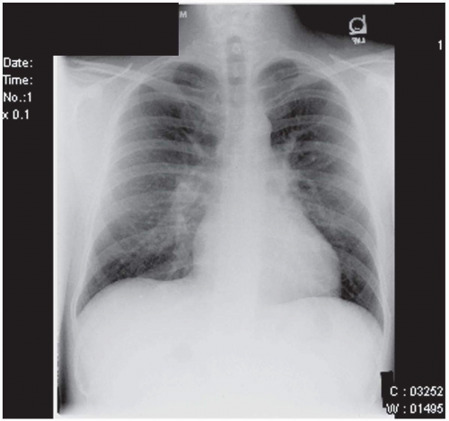


FIGURE 36.3. Chest radiograph of the same patient depicted in Fig. 36.2 with large secundum atrial septal defect, which demonstrates right ventricular enlargement and prominent peripheral pulmonary arteries, consistent with “shunt vascularity.”


FIGURE 36.4. Parasternal short-axis echocardiographic views of the right and left ventricles in systole and diastole, showing the septal motion typical of right ventricular volume overload in large ASD. The right ventricle is markedly dilated. In systole, the interventricular septum is normally positioned, and the left ventricle is round, which suggests that the left ventricular pressure exceeds the right ventricular pressure in systole. In diastole, the interventricular septum is bowed into the left ventricle, consistent with elevated right ventricular diastolic pressure.


FIGURE 36.5. Transesophageal echocardiography of secundum atrial septal defect in the horizontal and sagittal planes. On color Doppler echocardiography, a left-to-right shunt was demonstrated.


TABLE 36.2. Treatment, complications, and follow-up of adults with congenital heart disease


CONDITION


RECOMMENDATIONS
FOR REPAIR


COMMON
COMPLICATIONS


FOLLOW-UP


ENDOCARDITIS
PROPHYLAXIS


ASD


All primum ASD and sinus venosus if symptomatic and Qp/Qs ≥1.5:1 or


CHF
Atrial fibrillation


Every 2-5 yr after repair


No for secundum ASD and sinus venosus


Secundum ASD if asymptomatic with big RV


Yes before and after repair for primum ASD


VSD


Qp/Qs ≥1.5:1 and symptoms


CHF


Every 2-5 yr after repair


Yes before repair


Endocarditis


No 6 months after repair


Qp/Qs ≥2.0:1 without symptoms


Eisenmenger syndrome


Coarctation of the aorta


Upper extremity hypertension and gradient ≥20 mm Hg


Stroke
Aortic aneurysm
Aortic valve disease
Endocarditis and endarteritis


Yearly with MRI or CT of aorta every 2-5 yr


Yes


PDA


Audible murmur


Rare
Eisenmenger Syndrome


Periodically


Yes if audible


Rare endarteritis


No 6 months after closure


TOF


All affected adults without Eisenmenger syndrome who have not previously undergone repair
As needed for sequelae of operation


Pulmonary insufficiency
RV failure
Atrial and ventricular dysrhythmias


Yearly


Yes


ASD, atrial septal defect; CHF, congestive heart failure; CT, computed tomography; MRI, magnetic resonance imaging; PDA, patent ductus arteriosus; Qp/Qs, ratio of pulmonary blood flow to systemic blood flow; RV, right ventricle; TOF, tetralogy of Fallot; VSD, ventricular septal defect.


Therapy

Regardless of the patient’s age, closure of the secundum ASD improves symptoms and exercise tolerance and prevents the later development of congestive heart failure in
symptomatic patients with a pulmonary-to-systemic blood-flow ratio (Qp/Qs) of 1.5:1 or higher. Closure of the defect prolongs survival if performed before the age of 40 (10,11,14,15). ASD closure at an earlier age may prevent later occurrence of atrial fibrillation to some degree. However, atrial fibrillation is still frequent after repair and is associated with late stroke (10). Whether simultaneous performance of the Maze procedure for atrial fibrillation in the older patient with ASD prevents later arrhythmias is unknown. ASD closure is also recommended for asymptomatic patients younger than 40 years with a Qp/Qs of 1.5:1 or higher if right-sided heart enlargement is present, primarily for the prevention of right-sided heart failure. Closure of ASDs in asymptomatic older adults is more controversial. In one large study, 521 patients older than 40 years with secundum ASD were randomly assigned to undergo surgical closure or receive medical therapy and were monitored for more than 7 years (15). The risk of the combined end point (death, pulmonary embolism, major arrhythmic event, cerebrovascular embolic event, recurrent pulmonary infection, functional class deterioration, or development of heart failure) was higher in the medically managed group than in the surgical group (hazard ratio of 1.99; p < 0.01). Although no difference was found in mortality rate between medical and surgical groups during this period of follow-up, the authors propose surgical closure in all patients older than 40 with secundum ASD with a Qp/Qs of 1.7:1 or higher who do not have fixed pulmonary hypertension. Some patients with small secundum ASD and a history of paradoxic embolization may benefit from closure.

Indications for closure of sinus venosus and ostium primum ASD are less well defined. The higher incidence of mitral valve abnormalities in patients with ostium primum ASD and the higher risk of the development of pulmonary hypertension in patients with sinus venosus ASD suggest that most of these defects should be closed.

All studies of long-term outcomes after ASD repair have involved patients who underwent surgical closure through the standard surgical approach. Excellent short-and intermediate-term outcomes of percutaneous closure of ASDs with various devices have been reported (16,17,18). Currently two transcatheter atrial septal devices have been approved for clinical use in the United States (the CardioSEAL device, NMT Corporation, Boston, Massachusetts, and the AMPLATZER Septal Occluder, AGA Medical Corporation, Golden Valley, Minnesota). The CardioSEAL device is approved for closing the patent foramen ovale in individuals who have documented cerebral vascular accidents or other proven embolic events that are refractory to medical therapy, and the AMPLATZER Septal Occluder is approved for the closure of secundum ASD.

Individuals must have a secundum ASD or patent foramen ovale to be a candidate for device closure. Neither sinus venosus nor primum ASDs can be closed with any of these devices. Patients with defects with circumferential septal rims are the best candidates for closure. A patient is considered a candidate for device closure if the diameter of the ASD is less than 28 to 30 mm by transthoracic echocardiogram and if the diameter is less than 34 mm when stretched by a balloon catheter at the time of catheterization. In addition, the ASD must have a rim that is more than 4 mm away from all other cardiac structures, including the superior and inferior vena cavae, pulmonary veins, and the atrioventricular valves. Defects with multiple fenestrations can occasionally be closed with a single device, but multiple widely spaced defects are more difficult and require multiple devices.

A number of articles have established the safety and efficacy of the AMPLATZER device. One study reviewed the records of 89 patients who underwent either surgical closure (n = 44) or AMPLATZER device closure (n = 45) of an ASD performed between March 1998 and May 2000 (19). These investigators demonstrated that transcatheter closure with the AMPLATZER Septal Occluder carries the advantages of fewer complications, avoidance of cardioplegia and cardiopulmonary bypass, shorter hospitalization,
reduced need for blood products, and less patient discomfort. However, as with any other comparison between a surgical procedure and a transcatheter intervention, these authors acknowledged that the surgeon’s ability to close any ASD regardless of anatomy remains an important advantage of surgery. Similar results were reported in a multicenter nonrandomized study (20). The Amplatzer Septal Occluder has a low incidence of complications (21,22).

The experience from 13 European centers using the CardioSEAL double-umbrella devices to close interatrial communications in 334 patients has been reviewed (23). The investigators concluded that this device produced excellent results when used to close defects of small to moderate size. However, results are less optimal, and complications occurred when attempts were made to close very large defects. Late fractures of the arms of the CardioSeal device have limited its clinical utility. In summary, the AMPLATZER Septal Occluder is currently the device of choice for closing ASDs.

Percutaneous closure of ASDs eliminates scarring at the atrial suture line from surgery and, theoretically, may prevent atrial arrhythmias if performed at an early age. Also, minimally invasive, direct-access surgical techniques for ASD closure have been reported with no operative or late mortality and rare complications (24). Percutaneous treatment or minimally invasive surgery may be feasible for ASD closure in many patients.

According to the 2007 American Heart Association guidelines, bacterial endocarditis prophylaxis is not necessary for the preoperative patient with ASD. Endocarditis prophylaxis should be administered for 6 months after surgical or device closure (25).


Prognosis and Follow-up

In all adults with congenital heart disease, the physician must consider the complications of the lesion that has not been repaired (the natural history of the unrepaired defect), potential residua of incomplete repair, and sequelae of the operation. Patients with ASD who have undergone surgical closure in childhood, with either primary suture or patch closure, do well. El-Najdawi et al. (26) described long-term outcomes in 334 patients who underwent repair of primum ASD (partial atrioventricular septal defect) with or without mitral valve repair. Ten-, 20-, and 40-year survival rates were 93%, 87%, and 76%, respectively. Reoperation was required in 11% of patients, usually for mitral valve regurgitation or stenosis. When secundum ASD is repaired in childhood, operative mortality rates are less than 2%, and long-term survival is almost normal (27). In patients undergoing operation after 40 years of age, the operative mortality rate is slightly higher (5.8%) but still considered reasonable, and cardiovascular events may be less frequent than in medically treated patients (11.1% vs. 20.7%; hazard ratio, 2.0; p = 0.004) (15). Late atrial fibrillation is common and is associated with cerebrovascular events but is perhaps less frequent in patients undergoing operation before 40 years of age (10). Therefore, patients with ASD closed either in childhood or adulthood should not be considered “cured” but should be monitored for later development of arrhythmias and given anticoagulants if necessary.


VENTRICULAR SEPTAL DEFECT

Although ventricular septal defect (VSD) is the most common congenital heart defect diagnosed in childhood, it represents the third most common congenital cardiac anomaly in adults after ASD and patent ductus arteriosus (PDA) and accounts for 10% of congenital heart disease in adults. Spontaneous closure of small VSDs before adolescence (as occurs in approximately 60% of patients) (28), patient attrition due to congestive heart failure or arrhythmias in patients with large VSDs, and prior surgery are the reasons for the decreased incidence of VSD in adults. Locations of defects in the interventricular septum include perimembranous (beneath the crista supraventricularis when viewed from the right ventricle, and in the outflow tract of the left ventricle
beneath the aortic valve when viewed from the left ventricle) in 70%, muscular (either single or multiple in the muscular portion of the septum) in 20%, supracristal or subpulmonic (beneath the pulmonary valve, undermining the aortic valve annulus and associated with aortic valve prolapse and insufficiency) in 5%, and inlet, or “atrioventricular canal,” defects (beneath the septal leaflet of the tricuspid valve) in 5% to 8%.


Presenting Symptoms and Signs

The presentation of the adult with VSD is usually related to one of two extremes of very small or very large defects, inasmuch as most moderate or larger defects are diagnosed and surgically corrected in childhood. With small or “restrictive” VSDs, first described by Henri-Louis Roger in 1879 and given the moniker “maladie de Roger,” a large pressure difference exists between the left and right ventricle with a small left-to-right shunt and low pulmonary artery pressure. The affected adult patient is asymptomatic but often reports that a loud murmur was detected in childhood, perhaps during a sports physical examination. In rare cases, adults may be first seen with fever, septic emboli, and peripheral manifestations of endocarditis that result from infection of the “jet” lesion on the right ventricular free wall. The first and second heart sounds are normal. Typically, a loud, harsh grade III to IV/VI holosystolic murmur is auscultated in the third or fourth intercostal space at the left sternal margin, often associated with a palpable thrill. The murmur of the muscular VSD is very loud and harsh but may terminate abruptly in mid- to late systole as the defect is closed by myocardial contraction during systole. In some adults with supracristal VSD, a high-pitched diastolic blowing murmur of aortic insufficiency may be heard.

By adulthood, patients with large or “nonrestrictive” VSDs invariably progress to Eisenmenger syndrome with equilibration of right and left ventricular systolic pressures or development of suprasystemic pulmonary artery pressure with shunt reversal. Dyspnea is usually prominent, and exertional syncope is common. Occasionally, a patient may have a cerebrovascular event caused by paradoxic embolization or brain abscess from septic embolization. Development of a fever or new-onset neurologic symptoms should prompt investigation for a cerebral abscess. The patient may have cyanosis at rest or with exercise and usually exhibits clubbing of the fingers and toes. The jugular venous pressure is elevated. A right ventricular heave may be palpated at the left sternal border, and a prominent pulmonary artery impulse may be detected with or without a palpable pulmonic component of the second heart sound. A very loud P2 and right-sided S4 or S3 or both are common. Because shunting across the VSD is now minimal, the typical VSD murmur is absent. A holosystolic murmur of tricuspid regurgitation or a diastolic blowing murmur of pulmonary insufficiency (Graham Steele murmur) may be heard.

Occasionally, a moderate-sized VSD is first recognized in adults. In these patients, with defects usually less than half the size of the aortic orifice and a Qp/Qs of 2:1 or higher, symptoms include dyspnea, effort intolerance, and fatigue. If a history is found of a very loud murmur that has diminished or disappeared over the years, development of Eisenmenger physiology should be suspected. If aortic insufficiency has progressed, the diastolic murmur may be loudest, and physical findings of left-sided heart failure may be prominent with a displaced left ventricular impulse and a left-sided S3. In these patients, the holosystolic murmur of the VSD is generally softer than that in patients with small VSDs. If aortic insufficiency accompanies the VSD, the combined pansystolic and diastolic murmurs may be interpreted as a continuous murmur.


Helpful Tests

In patients with restrictive VSD, the ECG and chest radiograph are normal. The ECG is generally normal in the patient with moderate-sized VSDs, although it may reveal right axis deviation. The chest radiograph in
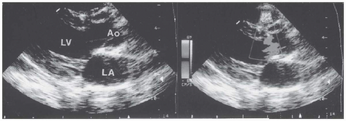
these patients may show cardiomegaly with enlarged left atrium and left ventricle (from the increased flow that returns from the lungs to the left side of the heart) and prominent central pulmonary arteries with “shunt vascularity.” In the patient with shunt reversal due to Eisenmenger physiology, the ECG shows prominent right axis shift with right atrial and right ventricular hypertrophy and possible right ventricular “strain” repolarization abnormality with QRS widening. Frequently, premature ventricular contractions are captured on routine ECG or ambulatory ECG monitoring. On chest radiograph, right-sided heart enlargement is notable, with dilated, sometimes calcified central pulmonary arteries and tapering or “pruning” of the pulmonary vessels in the periphery. On transthoracic color and spectral Doppler echocardiography, the hallmark of VSD is usually high-velocity flow from the left ventricle to the right ventricle at the site of the defect in the interventricular septum (Fig. 36.6). In some patients, a ventricular septal aneurysm forms as a result of partial or complete closure of a perimembranous VSD by portions of the septal leaflet of the tricuspid valve (Fig. 36.7). The sonographer must carefully interrogate all regions of the septum from multiple views with color and spectral Doppler imaging to avoid missing small defects. Nevertheless, very small muscular VSDs with characteristic murmurs, especially if multiple, may not be visualized on transthoracic or even the more sensitive transesophageal echocardiogram. Intravenous injection of agitated saline microbubbles is not usually helpful in the diagnosis of small VSDs, because the shunt is left to right. Adults with very large VSDs and Eisenmenger physiology may have no shunt whatsoever or a small right-to-left shunt demonstrated by color or spectral Doppler imaging at the defect site. Echocardiography is also instrumental for the evaluation of additional cardiac lesions, including aortic valve prolapse and insufficiency and subpulmonic obstruction, which may develop in late adolescence or adulthood as a result of hypertrophy of the crista supraventricularis. Cine magnetic resonance imaging and computed tomography may elegantly display all types of VSDs.


FIGURE 36.6. Transthoracic echocardiogram of a small perimembranous ventricular septal defect. Turbulence in the right ventricle, seen at the site of the defect on the color Doppler echocardiogram, resulted from the high-velocity left-to-right shunt.

In adults in whom closure of a moderate-sized VSD is under consideration, right- and left-sided heart catheterization may be necessary. Knowledge of pulmonary artery pressure and pulmonary vascular resistance is mandatory for making the decision for or against closure. In individuals with pulmonary arteriolar resistance that is two thirds of the systemic arteriolar resistance or more, lack of pulmonary vascular reactivity in response to pulmonary vasodilators such as oxygen or nitric oxide at the time of cardiac catheterization is predictive of high operative mortality risk. In rare cases, patients require open-lung biopsy to examine the
pulmonary vessels for evidence of irreversible pulmonary vascular obstruction.


FIGURE 36.7. Transesophageal echocardiogram of a ventricular septal aneurysm formed when the septal leaflet of the tricuspid valve became adherent to the perimembranous ventricular septal defect. No left-to-right shunt was visible on the color Doppler echocardiogram.


Differential Diagnosis

In the patient with restrictive VSD, the differential diagnosis includes other lesions that may produce a holosystolic murmur. Although the murmurs of mitral and tricuspid regurgitation are pansystolic, their locations (cardiac apex or lower left sternal border) and respiratory changes (augmentation of the tricuspid murmur with inspiration) help differentiate them from the murmur of VSD. Only rarely is the murmur of tricuspid or mitral regurgitation (e.g., with a flail leaflet) as harsh as that of VSD and associated with a palpable thrill. In patients with VSD and aortic insufficiency, the additive systolic and diastolic murmurs may be confused with the continuous murmur of the patent duct, ruptured sinus of Valsalva aneurysm, coronary artery fistula, or systemic-to-pulmonary artery collateral vessels such as those that occur with pulmonary atresia.


Complications

Until the mid-1990s, bacterial endocarditis was thought to be the only significant potential complication of the restrictive VSD, with incidences of 1.9 per 1,000 patient years in unrepaired simple VSD and 3.5 per 1,000 patient years if aortic regurgitation coexisted with the VSD (29). The incidence of endocarditis after closure of the VSD decreases to 0.75 per 1,000 patient years. However, in a study of 188 adults (aged 17 to 72) with small unrepaired VSDs, 26.6% of whom had additional cardiac lesions, such as bicuspid aortic valve and coarctation of the aorta, complications occurred in 53% of patients (30). Of note, spontaneous
closure of the VSD occurred in 19 patients (10%, all between the ages of 17 and 45; mean age, 27 years). Serious complications occurred in 46 patients (25%): infective endocarditis in 11%, progressive aortic insufficiency in 5%, and symptomatic arrhythmias, most commonly atrial fibrillation, in 8.5%. Irreversible pulmonary vascular obstruction (Eisenmenger syndrome) inexorably develops before adulthood in the patient with a large, nonrestrictive VSD and carries the potential for complications such as endocarditis; right-sided heart failure; hemoptysis; erythrocytosis with the attendant risks of hyperviscosity, hyperuricemia, and gout; renal insufficiency; cholelithiasis; and sudden cardiac death. Maternal and fetal mortality rates among pregnant women with Eisenmenger syndrome exceed 50% and are higher in women with pulmonary hypertension due to VSD than with that due to ASD (31). Aortic insufficiency develops in 2% to 7% of patients with VSDs over time, occurs more commonly in adults older than 20 years, and is classically associated with the supracristal or subpulmonic type of defect, although it may occur in perimembranous VSD (32). In 25% of these patients in one study, the severity of aortic insufficiency remained stable over time. Aortic insufficiency is much more common in the Asian population with subarterial VSDs, developing in 64% of 139 asymptomatic patients in one study (33). The authors of this study found that aortic prolapse and insufficiency developed only in patients with a VSD diameter larger than 5 mm and recommended closure in these patients.


Therapy

Historically, the only recommended therapy for patients with restrictive VSD is bacterial endocarditis prophylaxis for dental and other procedures. The most recent American Heart Association guidelines no longer recommend endocarditis prophylaxis for this population (25). With future advances in the technique of percutaneous closure of VSDs, it is possible that the risk accompanying the procedure will one day be lower than the risk of complications in these patients, and closure may eventually be recommended.

Surgical closure is recommended for adults with a Qp/Qs of 1.5:1 or higher with symptoms, evidence of left ventricular enlargement or dysfunction, and pulmonary hypertension that is not severe. If pulmonary vascular resistance is normal, closure of defects with a Qp/Qs of 2:1 or higher in the asymptomatic patient is probably warranted to prevent the progression to pulmonary hypertension, because the risk of death is 4 times higher in these patients (34). Patients with severe pulmonary hypertension (pulmonary-to-systemic vascular resistance ratio higher than 2:3) who have a net left-to-right shunt flow ratio of 1.5 or higher may safely undergo repair only if reversibility of pulmonary resistance can be demonstrated at catheterization or by lack of permanent vascular obstruction on lung biopsy. Because aortic insufficiency is progressive in patients with supracristal VSD, closure seems warranted if insufficiency is more than mild or is shown on serial echocardiography to be progressive. A history of endocarditis, especially if recurrent, may also be an indication for closure.

Studies of percutaneous transcatheter closure of VSDs have, to this time, included a limited number of patients with fairly brief follow-up (35). A recent multicenter study of transcatheter closure of perimembranous ventricular septal defect(100 patients, mean age 9 years), demonstrated successful deployment in 93% of patients. Complications occurred is 29% of patients, including complete heart block in two patients and new or increased aortic or tricuspid regurgitation in 18 patients. Complete closure was present in 58% of patients immediately after closure, and in 83% of patients 6 months after closure. Given the high success rate and low morbidity and mortality of surgical closure of VSD, further development and study of these devices before their widespread use can be advocated. As with ASDs, a minimally invasive surgical approach to closure of VSDs is used at some centers (36).

Patients with fixed pulmonary vascular obstruction have a very high mortality rate
with repair and should not undergo operation. At this time, no curative therapy is available for patients with Eisenmenger syndrome. Pregnancy should be strongly discouraged, and sterilization of the patient or, preferably, the sexual partner should be offered, because even minor noncardiac surgery conveys significant risk in these patients. Pregnancy termination may be considered for certain patients.

Continuous intravenous prostacyclin may improve hemodynamics and quality of life in symptomatic patients with pulmonary hypertension and congenital heart disease (37). In 20 such patients receiving long-term prostacyclin therapy, mean pulmonary artery pressure decreased by 21%, cardiac index improved from 3.5 ± 2.0 L per minute per square meter to 5.9 ± 2.7 L/minute/m2, and New York Heart Association functional class improved from 3.2 ± 0.7 to 2.0 ± 0.9 (p < 0.0001). A recent randomized, placebo-controlled, multicenter trial of 37 patients with Eisenmenger syndrome due to a variety of congenital heart defects demonstrated improvement in 6-minute walk distance of 53 meters (38). No increased risk of adverse events was found as compared with the placebo group, and no significant decrease in oxygen saturation. The treatment duration was relatively short, at 16 weeks. Interest has been expressed in the use of sildenafil and related agents for this patient population. A trial of 20 patients, including 10 with idiopathic pulmonary hypertension and 10 with Eisenmenger syndrome, demonstrated significant improvement in 6-minute walk and decrease in pulmonary artery pressure as compared with placebo (39). Surgical treatment of patients with Eisenmenger syndrome either with heart-lung transplantation or with single- or double-lung transplantation and concomitant intracardiac repair has been performed in a limited number of patients, with a disappointing 1-year survival rate of less than 60% (40,41).

All patients with unrepaired VSD and Eisenmenger syndrome should receive endocarditis prophylaxis. Residual defects after surgical repair require lifelong prophylaxis. Patients with native, unrepaired VSDs, and those more than 6 months after surgical closure do not require endocarditis prophylaxis (25).


Prognosis and Follow-up

As noted previously, the course of unrepaired patients with small VSDs may not be as uneventful as originally thought; 25% of patients eventually experience endocarditis, progressive aortic regurgitation, left ventricular dysfunction, or atrial arrhythmias. However, no significant difference in survival occurred from that of the normal population in the study (30). In patients with unrepaired VSD and Eisenmenger syndrome, survival rate is 77% at 15 years of age but only 42% at 25 years of age. Death occurs suddenly in 30% of patients and is due to congestive heart failure in 25%, hemoptysis in 15%, and pregnancy, noncardiac surgery, and endocarditis in the remainder (42).

The life expectancy of patients with VSD and normal pulmonary vascular resistance repaired in childhood is nearly normal. Of 296 patients who underwent surgical closure during the early operative era of 1954 to 1961, the survival rate was 80% at a mean of 26.8 years of follow-up (43). Mortality rates were higher in patients who underwent operation after the age of 5 years, in those with elevated pulmonary vascular resistance higher than 7 Woods units, and in those with transient or permanent complete heart block. Complete heart block infrequently occurs late after VSD repair and is not less frequent after atriotomy than after a ventricular incision (44). Atrial fibrillation occurs in approximately 8% of patients after repair, and the frequency increases at older ages (30). Ventricular arrhythmias are common but generally benign; however, a higher incidence of sudden death has been reported in patients with surgically managed VSDs than in those with medically managed VSDs, presumably from ventricular arrhythmias (34).

Only gold members can continue reading. Log In or Register to continue

Stay updated, free articles. Join our Telegram channel

Aug 18, 2016 | Posted by in CARDIOLOGY | Comments Off on Congenital Heart Disease in Adults

Full access? Get Clinical Tree

Get Clinical Tree app for offline access