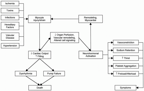
Heart Failure Due to Left Ventricular Systolic Dysfunction


Heart Failure Due to Left Ventricular Systolic Dysfunction


David B. Dyke

Todd M. Koelling


Heart failure is a clinical syndrome in which any abnormality of cardiac function causes either a failure of the heart to pump blood at a rate commensurate with the requirement of metabolizing tissues, or a situation in which filling pressures are elevated, or frequently both conditions simultaneously. Patients with impaired cardiac-pumping function experience symptoms related to abnormal perfusion and retention of vascular fluid volume. The cardinal symptoms of heart failure include fatigue or exercise intolerance, dyspnea, and edema, although other related symptoms may also occur. Heart failure may be caused by disorders of the pericardium, myocardium, heart valves, or great vessels, but most patients manifest the syndrome through abnormalities in systolic function. Reduction in myocardial contractility is more commonly referred to as systolic dysfunction and may also coexist with chamberfilling abnormalities, also referred to as diastolic dysfunction. This chapter focuses on patients with heart failure due to systolic dysfunction.

The term heart failure is now preferred to congestive heart failure, inasmuch as not all patients with heart failure are “congested,” and experts believe that the latter description has limited diagnostic accuracy. Heart failure occurs commonly in clinical practice and represents the most common Diagnosis-Related Group discharge diagnosis in the Medicare (elderly) population. Nearly 5,200,000 patients (2,600,000 men and 2,600,000 women) in the United States have heart failure, and approximately 550,000 new cases of heart failure are diagnosed each year. Of Americans older than age 60, approximately 5.2% to 12.4% have experienced heart failure. Although heart failure can occur at any age, most patients with heart failure are older than 65 years (1).

Heart failure is a common cause of mortality in the population; nearly 300,0000 patients die of heart failure each year. Over the period from 1994 to 2004, deaths from heart failure increased by 28%. Since 1979, hospitalizations for heart failure as a primary diagnosis have increased by 175% (399,000 in 1979 to 1,099,000 in 2004) (Fig. 14.1). The annual estimated rate of new and recurrent heart-failure events for white men aged 65 to 74 is 15.2 per 1,000 population; for those aged 75 to 84, it is 31.7; and for those aged 85 and older, it is 65.2. For black men, the rates are 16.9, 25.5, and 50.6, respectively. For white women in the same age groups, the rates are 8.2, 19.8, and 45.6, respectively, and for black women, the rates are 14.2, 25.5, and 44.0, respectively.

Because heart failure necessitates frequent hospitalizations in the population, the costs of caring for this syndrome are considerable. The estimated direct and
indirect cost of heart failure in the United States for 2007 is $33.2 billion dollars (1).


FIGURE 14.1. Hospital discharges in the United States by gender from 1979 to 2004. (From: Centers for Disease Control and Prevention/National Center for Health Statistics and the American Heart Association, with permission.)


USUAL CAUSES

It has been well recognized that the myocardium undergoes structural changes in response to cardiovascular disease states. Irrespective of the initial cause, myocardial damage results in reduced power output of the heart, which occurs in a varying time frame, depending on the severity of disease and the cause of myocardial damage. Myocardial ischemia is usually manifested initially by regional impairment of myocardial function. Nonischemic dilated cardiomyopathies caused by such insults as viral infections, exogenous toxins, regurgitant valvular lesions, and hereditary factors normally manifest with global left ventricular dysfunction. Abnormalities that begin by causing elevated myocardial strain include hypertension, aortic stenosis, and hypertrophic cardiomyopathy. Most patients with these abnormalities are first seen with myocardial hypertrophy, either concentric (hypertension, aortic stenosis) or focal (hypertrophic cardiomyopathy). Although the types of insults that occur may be very different in the initial appearance of the myocardium, the chronic adaptations that occur in the body as a response to myocardial dysfunction reach a common pathway, as it is understood today (Table 14.1).


Specific Etiologies

The term cardiomyopathy is an ill-described, general category for a large group of unrelated disease processes that share only the clinical characteristic of substantially reduced cardiac pumping function and power output. Practical and graphic descriptions have been used to describe cardiomyopathy, and this classification is firmly anchored in the pathologic description of the heart. Thus dilated cardiomyopathy, hypertrophic cardiomyopathy, and infiltrative cardiomyopathy are descriptive pathologic terms. Alternatively, cardiomyopathy is characterized by the specific clinical or disease process with which it is associated. Thus terms such as peripartum cardiomyopathy, diabetic cardiomyopathy, and toxic cardiomyopathy are used. In clinical practice, the etiology of left ventricular systolic dysfunction can be identified in patients with coronary artery ischemia or infarction, infectious vectors, toxins, hereditary conditions, and conditions for which no cause can be identified (idiopathic) (Table 14.1). These disorders, in aggregate,
account for the majority of cases of heart failure resulting from myocardial disease.


TABLE 14.1. Causes of systolic heart failure


Primary myocardial diseases


Inherited (genetic) cardiomyopathic disorders


Dilated cardiomyopathy


Late remodeling stage of hypertrophic cardiomyopathy


Arrhythmogenic (right) ventricular cardiomyopathy


Left ventricular noncompaction


Muscular dystrophies


Secondary myocardial diseases


Energy-supply deficit


Connective tissue diseases


Coronary atherosclerosis


Systemic lupus erythematosus


Coronary dissection


Infiltrative diseases


Coronary embolus


Sarcoidosis


Excess ventricular afterload


Amyloidosis


Hypertension


Hemochromatosis


Aortic stenosis


Wilson disease


Aortic coarctation


Toxins


Excess ventricular preload


Alcohol, cocaine


Mitral regurgitation, tricuspid regurgitation


Anthracycline


Aortic insufficiency, pulmonic insufficiency


Doxorubicin


Tachycardia mediated


Daunorubicin


Atrial fibrillation/flutter


Epirubicin


Supraventricular tachycardia


Paclitaxel


Infectious


Trastuzumab (Herceptin)


Viral myocarditis


Cyclophosphamide


Postrheumatic


Interferon


Septicemia related


Interleukin-2


Human immunodeficiency virus


Chloroquine


Protozoal (Trypanosoma cruzi)


Zidovudine


Endocrine


Thoracic radiation therapy


Hypothyroidism, hyperthyroidism


Congenital heart disease


Pheochromocytoma


Peripartum cardiomyopathy


Diabetic


Stress-induced “Tako-tsubo” cardiomyopathy


Obesity related


Idiopathic cardiomyopathy


Ischemia

In patients with coronary artery disease, a cardiomyopathy can develop as the result of one extensive myocardial infarction, multiple smaller myocardial infarctions, ongoing ischemia from severe triple-vessel disease, or coronary artery disease associated with significant mitral regurgitation. Myocardial dysfunction may also develop after coronary artery bypass surgery, even in the setting of otherwise technically adequate graft placement. Early identification of myocardial dysfunction associated with coronary artery disease is important, in view of the potential for reversal of dysfunction with effective management. When overt angina is not apparent, because of limited exercise capacity, subclinical myocardial ischemia may nonetheless produce abnormal systolic and diastolic ventricular dysfunction. Additional subclinical loss of viable myocardium also may occur.

When ischemia-related myocardial dysfunction prevents identification of reversible disease during exercise, viable hibernating
myocardium may be identified by nuclear imaging studies. The predictive value of 201T1 and 99mTc scintigraphy for detecting hibernating myocardium has been enhanced by newer redistribution and reinjection protocols, and these techniques are more readily available than is positron-emission testing at most institutions. In addition, newer techniques using magnetic resonance imaging also are being used for diagnosis of viable myocardium. Once identified, reversible ischemia caused by hibernation can be managed with interventional or surgical techniques, to prevent further deterioration of myocardial function.


Infections

A long list of infectious agents can be identified for this subgroup of myocardial disease. Myocardial dysfunction develops from a nonspecific immune or inflammatory response, or both, or from structural damage to cardiac myocytes. “Viral” myocarditis is frequently suspected among the patients who are otherwise classified as having idiopathic cardiomyopathy, although many of these patients may actually have an inherited cardiomyopathy. Myocarditis can be diagnosed with endomyocardial biopsy and has been shown to be present in 12% of patients seen with dilated cardiomyopathy in the absence of coronary artery disease within 6 months of their original diagnosis (2). With the exception of a few welldescribed viral causes that can be inferred from serial immune titers, isolation of a specific viral vector remains difficult. Molecular biologic techniques permit enhancement of viral messenger ribonucleic acid. However, in evaluations of myocardial tissue by histologic techniques, the occurrence of viral particles is equally distributed between patients with active myocarditis and those with nonspecific cardiomyopathy. Etiologic origin is therefore difficult to assign.

Diagnosing and treating active myocarditis remains a challenge. In general, the diagnosis can be confirmed only on myocardial biopsy, and the occurrence may be sporadic and related to fluctuation in Coxsackie virus prevalence. Endomyocardial biopsy also can be used for histologic documentation when a strong clinical suspicion of myocarditis is present. Endomyocardial biopsy evidence of myocarditis, on the whole, carries a prognosis similar to that for heart failure of idiopathic origin.

Among patients with history of a flulike syndrome in the setting of clinical suspicion of myocarditis, biopsy evidence of inflammatory infiltrates is seen in approximately 25%, and yet almost half of these patients may have other concurrent disorders. Investigators have shown that the shorter the duration of illness, the greater the likelihood of a biopsy sample positive for inflammation; the likelihood reaches almost 90% in patients seen within 4 weeks of their initial symptoms (3). Seasonal and yearly variations exist in the clinical presentation. The Myocarditis Treatment Trial demonstrated a better-thanexpected prognosis in patients with myocarditis (4). Specific immunosuppression did not alter outcome. Additional analyses are in progress to determine immunologic markers that identify patients who may benefit from immunosuppressive therapy.

Myocarditis may be present in cases in which the biopsy result is negative. One possible explanation is that the transvenous technique does not sample enough myocardial sites to detect each case of a disease that may have a focal or multifocal distribution. Acute myocarditis may occur as a regional process and can mimic acute myocardial infarction, with striking electrocardiographic changes. The regional nature of this disorder is evident with invasive and noninvasive assessment of ventricular performance. Depending on clinical presentation, this disorder may necessitate cardiac catheterization, which is the only means of definitively excluding epicardial coronary artery disease.

Myocardial dysfunction due to Chagas disease remains the most common worldwide cause of cardiomyopathy (5). Infection in humans is caused by a bite from the reduviid insect, which harbors the protozoa Trypanosoma cruzi in its gastrointestinal tract. T. cruzi is the infectious etiology of Chagas disease and gains entry into a human host by fecal deposition after a bite
from the reduviid. After initial infection, acute trypanosomiasis occurs, followed by a long latent period; chronic Chagas disease appears up to 20 years later. Myocardial dysfunction and congestive heart failure develop during this time. T. cruzi parasites within a cellular infiltrate may be present during the acute phase, but cardiac manifestations occur during the chronic phase. No correlation is found between the severity of disease and parasitemia. T lymphocytes may destroy normal myocardial cells, and antibody-mediated responses to specific myocyte components, such as the sarcoplasmic reticulum, have been identified.

In the Western Hemisphere, the greatest concentration of this disorder is in Central and South America, where 20 million people may be infected with the parasite (5). However, with increasing migration from these regions to the United States, consideration must be given to this diagnosis in patients of Latin American or South American origin from endemic regions. The concern regarding Chagas disease as an etiology of myocardial dysfunction is not limited to the endemic populations. Currently, donor blood products are not routinely screened for T. cruzi. Thus blood donation is conceivably a means by which T. cruzi may be spread by a nonvector pathway. An acceptable method to screen blood products routinely for T. cruzi has not been established. This has not proved to be a significant problem at the current time, but as much as 10 to 15 years may separate acute parasitemia and overt myocardial dysfunction.


Toxins

Several exogenous toxins are well known to cause left ventricular systolic dysfunction and subsequent heart failure. The most common of these is alcohol, represented in 3.4% of cases of systolic heart failure in the absence of coronary artery disease. Myocardial toxicity due to anthracyclines (e.g., doxorubicin) is a common cause of systolic heart failure in patients who received chemotherapy for the treatment of cancer (6). Other exogenous toxins known to lead to systolic heart failure include cocaine, other chemotherapeutic agents (cyclophosphamide and trastuzumab), interferon, interleukin-2, and chloroquine. In the mid-1960s, a clustering of cases of acute-onset cardiomyopathy developing in patients with heavy beer consumption led to the discovery that cobalt represented a myocardial toxin. When the practice of adding cobalt to beer was stopped, no further cases occurred.


Alcoholic cardiomyopathy

Alcohol may be associated with heart failure in several different ways. Alcohol causes an acute depressant effect on myocardial contractility that can result in measurable dysfunction with binge drinking. Evidence suggests that the fundamental mechanism of injury induced by ethanol is structural and chemical disorganization of membranes, interference with ion transport, and derangement of various biochemical functions that possibly allow calcium to accumulate in the cell (7). Heavy alcohol consumption may also cause atrial tachyarrhythmias, termed holiday heart, which may contribute to the development of systolic dysfunction. The amount of alcohol necessary to cause this is unknown, because the testimony of alcoholic patients regarding intake cannot be validated. Studies have shown that ejection fraction correlates inversely with reported alcohol intake in alcoholic patients, and women may be more sensitive to the myocardial toxicity of alcohol than are men.

The pathologic and physiologic characteristics of alcoholic cardiomyopathy are similar to those of idiopathic dilated cardiomyopathy in gross appearance. The morphometric evaluation of endomyocardial biopsy does not provide adjunctive prognostic information in these patients. As many as one fourth of patients with systolic failure due to alcohol may have elevated cardiac output, caused by concomitant liver disease and development of arteriovenous fistulae. Patients with alcoholic cardiomyopathy may have a favorable prognosis in comparison with those with idiopathic cardiomyopathy; approximately 50% of alcoholic patients experience improved
left ventricular function once abstinence is established.


Athracycline and anticancer-related cardiomyopathy

Doxorubicin and daunorubicin are anthracycline analogues that are widely used as chemotherapeutic agents. One important side effect caused by anthracyclines is cardiotoxicity. Cardiotoxicity due to anthracycline derivatives has been shown to be dependent on the cumulative dose (6). Measurable left ventricular systolic dysfunction is rare in patients receiving less than 350 mg per square meter of doxorubicin but may be seen in as many as 30% of patients receiving more than 600 mg per square meter (8). The peak levels of the drug may be a determinant for developing the disorder: Some evidence suggests that giving the same total dose weekly rather than every 3 weeks or administering the drug by slow continuous infusion rather than by bolus may reduce the incidence of cardiotoxicity. The risk factors for development of doxorubicin-induced cardiomyopathy include age older than 70, use in combination with other chemotherapeutic agents, concomitant or prior mediastinal radiotherapy, prior cardiac diseases, hypertension, liver disease, and whole-body hyperthermia. Most authors on this topic have recommended monitoring patients serially with radionuclide ventriculography as patients are treated with anthracyclines (6). The diagnostic test with the greatest specificity and sensitivity for doxorubicin-induced cardiomyopathy is endomyocardial biopsy. Endomyocardial tissue from the right ventricle shows typical histopathologic changes, including loss of myofibrils, distention of the sarcoplasmic reticulum, and vacuolization of the cytoplasm; these may appear before measurable changes in left ventricular systolic function occur. A biopsy scoring system has been described to show that in patients in whom more than 25% of cells exhibit histopathologic changes, substantial changes in the ejection fraction will probably develop, which suggests that treatment should be terminated (9). However, it is still possible that in patients with lower biopsy grades, cardiomyopathy will develop 4 to 20 years later.

In addition to toxicity related to anthracyclines, several other anticancer therapeutics are associated with development of cardiomyopathy, the most common of which is trastuzumab (Herceptin), used for the treatment of breast cancer. The frequent use of multiple anticancer therapies (including radiation therapy) can greatly increase the future likelihood of developing cardiomyopathy.


Hereditary Influences

Studies have shed light on the role of the genetic background in the onset and the development of heart failure due to diastolic dysfunction (hypertrophic disease) and systolic dysfunction. Familial forms of dilated cardiomyopathy are common and have been described in as many as 30% of patients with nonischemic cardiomyopathy. This condition appears genetically highly heterogeneous. Inheritance patterns are usually autosomal dominant, but X-linked, autosomal recessive, and mitochondrial inheritance have been described as well. Genetic abnormalities found to be associated with familial dilated cardiomyopathy include mutations in genes encoding cytoskeletal/sarcolemmal, sarcomere, nuclear envelope, and transcriptional coactivator proteins. It has been postulated that the molecular defects involved in hereditary forms of dilated cardiomyopathy cause an abnormality in the transmission of contractile force. Polymorphisms in the Ile164 β2-adrenergic receptor have been shown to be associated with poor prognosis in patients with dilated cardiomyopathy (10). In a study of 259 patients with dilated cardiomyopathy and NYHA classes II to IV symptoms, patients with the Ile164 polymorphism displayed a striking difference in survival rates, with a relative risk of death or need for cardiac transplantation of 4.81. Further studies of the genetic determinants of dilated cardiomyopathy should allow
better understanding of the underlying mechanisms that promote the progression of the disease, to identify subjects at risk of the disease who would benefit from early medical management and to promote the development of pharmacogenetics.


Idiopathic Causes

Idiopathic remains the designation for many forms of dilated cardiomyopathy, when coronary artery disease and specific causes such as those listed earlier have been excluded. With the exception of primary causes, which are clinically identified, limited screening procedures can identify a specific cause. Of these, perhaps the most fruitful is the screen test for thyroid disease, which may be particularly important in the evaluation of the elderly patient. Both hyperthyroidism and hypothyroidism may produce left ventricular dysfunction. Abnormalities of trace substances such as selenium have been suggested, but deficiencies do not routinely occur in Western diets. Patients may also have left ventricular systolic dysfunction due to chronic tachycardia conditions, such as atrial fibrillation with rapid ventricular response. Rate control with β-blockers, digitalis, or both has been shown to lead to improvements in left ventricular function on follow-up testing.


BIOLOGIC MECHANISMS AND DISEASE PROGRESSION

Occurrence of the primary disorder leads first to myocyte injury or increased myocyte strain, or both (Fig. 14.2); second, to myocardial remodeling in structure and function; and, third, to loss of systolic or diastolic function, which in turn leads to decreased cardiac output and elevated filling pressures. Structural changes in the myocardium and vasculature are important contributors to the progression of left ventricular dysfunction. Myocardial fibroblasts and vascular smooth muscle cells may hypertrophy or proliferate, or both, in response to a variety of stimuli. These structural effects lead to changes in the compliance of arteries that augment the left ventricular load and to increases in the volume or mass (or both) of the left ventricle (11). The role of such ventricular remodeling in heart failure has been further described in patients who have had myocardial infarctions.

Many of the adaptive and maladaptive responses in congestive heart failure occur at sites distal to the initial myocardial damage. Decrements in cardiac output and elevations in central venous pressure result in reduced organ perfusion. Underperfusion of the kidney and underfilling of the arterial vasculature result in a cascade of adaptations that lead to neurohormonal alterations that have been found to have direct myocardial toxicity through myocyte hypertrophy, myocardial fibrosis, apoptosis, or a combination of these (12). Nonetheless, these distal abnormalities are integrally related to reduction of systolic function. Because cardiac myocytes cannot replicate at a rate sufficient to contribute to repair a direct injury, the response to injury is limited primarily to hypertrophy and increased interstitial tissue alterations.

The mechanisms that lead to progression of disease in heart failure include neural and hormonal factors that increase the load on the left ventricle, stimulate growth of myocytes, and may have direct toxic effects on the myocardium. The concentrations of several neurohormones and cytokines, including plasma norepinephrine, plasma renin activity, atrial natriuretic peptide (ANP), and tumor necrosis factor, have been shown to be increased in plasma in patients with congestive heart failure. The elevation in the levels of these compounds becomes more marked as clinical symptoms of heart failure advance and is associated with increased mortality rates. Evidence also indicates that elevations of these compounds may be a more sensitive method of monitoring disease progression, as Benedict et al. (13) showed in the Studies of Left Ventricular Dysfunction (SOLVD) prevention trial that plasma norepinephrine levels continued to be
predictive of mortality and development of clinical events related to the onset of heart failure, despite the patients’ being asymptomatic or minimally symptomatic.


FIGURE 14.2. Common mechanisms in disease progression in patients with heart failure.


Renin-Angiotensin-Aldosterone System

Activation of the renin-angiotensin-aldosterone system is one of the predominant abnormalities of heart failure. The degree of increase in plasma renin activity provides an indicator for prognosis in patients with heart failure (14). Studies in mild and asymptomatic heart failure demonstrate relatively less activation, but even these values are increased in comparison with normal. The degree of renin activity is intensified in the presence of diuretic therapy. Angiotensin II causes constriction of the systemic vasculature and vasoconstriction of both the afferent and efferent renal arterioles. In some patients with severe heart failure, treatment with angiotensin-converting enzyme (ACE) inhibitors may cause a deterioration of renal function. This may be related to fixed renal artery disease or, alternatively, to selective blocking of the constrictor action of angiotensin II on the efferent arteriole (15). Renin-system components have been identified in the myocardium and vasculature, where they adversely affect fibrosis and remodeling, as well as cellular dysfunction. These findings suggest that the renin-angiotensin-aldosterone system has effects on cardiac function beyond altering sodium excretion and cardiac afterload. Not only is angiotensin II a potent vasoconstrictor, but it also causes a direct effect on hypertrophy of myocytes and may lead to energy-supply mismatch as the capillary bed perfuses a larger bed.

In addition to vasoconstriction, angiotensin II stimulates aldosterone secretion by the adrenal gland, producing sodium retention and potassium excretion at the distal nephron. Elevations in the activity of aldosterone lead to a sodiumretentive state found in patients with heart failure. Although adrenergic stimulation and angiotensin II increase sodium transport in the proximal tubule of the kidney, patients with increased activity from aldosterone overcome this effect, and sodium delivery to the distal tubules is attenuated, leading to the edematous state. It was shown previously that although ACE inhibition continues to suppress angiotensin II levels over the course of 1 year, levels of aldosterone are initially suppressed during the first 1 to 3 months of therapy but fail to be suppressed beyond 6 months of therapy (16). This is thought to occur because stimuli in the form of glucocorticoids, hyperkalemia, hypermagnesemia, melanocytestimulating hormone, and endothelin continue to increase aldosterone secretion, although angiotensin II levels are low. Analysis of the Cooperative North Scandinavian Enalapril Survival Study (CONSEN-SUS) revealed that elevated levels of aldosterone were associated with the lowest rates of survival, and a reduction in plasma aldosterone during the course of therapy was associated with a favorable impact on survival (17). Although elevated aldosterone levels may track the clinical state of patients with congestive heart failure, they may also be responsible in part for progression of myocardial dysfunction through mechanisms that lead to abnormal accumulation of collagen, which surrounds and encases myocytes, resulting in diastolic and systolic ventricular dysfunction. Such deposition of collagen may lead to pathologic hypertrophy of the myocardium and has been shown to be prevented by spironolactone in a rat model of arterial hypertension (18). Spironolactone at doses of 25 to 50 mg per day can be used safely in conjunction with ACE inhibitors, diuretics, and digitalis. In a minority of patients, however, hyperkalemia may occur, leading to discontinuation of the drug. Findings of the Randomized Aldactone Evaluation Study (RALES) revealed that doses of spironolactone as low as 12.5 mg per day significantly reduce atrial natriuretic factor levels in patients with classes II to IV heart failure without a significant effect on serum potassium levels (19). Moreover, the RALES investigators also found that treatment groups had lower levels of aldosterone, norepinephrine, and plasma renin activity.


Sympathetic Nervous System

The baroreceptor-mediated increase in sympathetic tone that occurs with ventricular dysfunction has several consequences, including increased myocardial contractility, tachycardia, arterial vasoconstriction and thus increased cardiac afterload, and venoconstriction with increased cardiac preload. β-Adrenergic receptors in the heart either are downregulated (β1-adrenegic receptors) or have abnormalities in signal-transduction activity that effectively uncouple them from effector mechanisms (β1– and β2-adrenergic receptors) (15). Increased local and circulating concentrations of norepinephrine may contribute to myocyte hypertrophy, either directly through stimulation of α1– and β-adrenergic receptors or secondarily by activating the renin-angiotensin-aldosterone system. Norepinephrine is directly toxic to myocardial cells, an effect mediated through calcium overload, the induction of apoptosis, or both. Norepinephrine-induced death of myocytes can be prevented by concomitant nonselective β-adrenergic blockade. Patients with plasma norepinephrine concentrations greater than 800 pg per milliliter (4.7 nM) have a 1-year survival rate of less than 40% (20). Through renal vasoconstriction, stimulation of the renin-angiotensin-aldosterone system, and direct effects on the proximal convoluted tubule, increased renal adrenergic activity contributes to the avid renal sodium and water retention that occurs in patients with heart failure.

Substantiating the importance of the sympathetic nervous system in the heart failure syndrome, multiple randomized controlled trials have demonstrated the benefits of β-adrenergic blockade on clinical outcomes in patients with heart failure (21,22,23). In the past, β-adrenergic blockade was thought to be contraindicated in patients with heart failure. However, if patients can tolerate short-term β-adrenergic blockade, ventricular function subsequently improves.


Natriuretic Peptides

The roles of atrial natriuretic peptide (ANP) and brain natriuretic peptide (or B-type natriuretic; BNP) in congestive heart failure and mitral regurgitation are not well understood. Investigators have shown that these compounds are produced by cardiac myocytes and their levels correlate inversely with left ventricular ejection fraction, directly with left atrial pressure, and directly with New York Heart Association (NYHA) class and mortality (24). Administration of exogenous ANP, 0.10 µg per kilogram per minute to normal subjects, was found to increase sodium (450%) and free water (100%) excretion, while decreasing plasma renin (33%) and aldosterone (40%). Similar administration of ANP to patients with congestive heart failure had no effect on sodium or free-water excretion, although significant decreases in pulmonary capillary wedge pressure (19%), systemic vascular resistance (13%), and plasma aldosterone (51%), and increases of cardiac index (17%) were noted. More recently, nesiritide, an intravenous form of BNP, has been shown to improve symptoms and reduce elevated filling pressures in patients treated for heart failure in various hospital settings (25). Although use of nesiritide has become routine in clinical practice, concern over long-term safety of this drug has surfaced as a result of recently published meta-analyses of several randomized clinical trials demonstrating possible decrements in both renal function and survival (26,27). Results of an ongoing randomized, placebo-controlled mortality trial should help us understand better the potential benefit of the use of nesiritide in patients with heart failure.

Serum level of endogenous BNP has been shown to be an independent prognostic indicator in patients with cardiac disease. Because of this, point-of-care BNP testing has also become part of routine clinical practice, and although it has been demonstrated to be helpful for aiding in establishing a diagnosis in patients with new-onset dyspnea (28), the value of serial measurements of BNP to guide therapy for patients with established heart failure is not well established.


Endothelin

Endothelin (ET) is a family of potent vasoconstrictor peptides of vascular endothelial
origin. Although it has been proposed that the vasoconstrictor effects of ET are produced at the local vascular level, increased plasma concentration of ET has been identified in cardiovascular disorders (29). ET levels have been demonstrated to be nearly threefold higher in patients with congestive heart failure than in normal controls. ET was used a decade ago as a potent vasoconstrictor. The peptides were originally identified from rodent sources (ET-3) as well as human and porcine sources (ET-1). ET-1, ET-2, and ET-3 all have potent vasoconstrictor properties. ET-1 levels have a close association with pulmonary pressures, as well as the resistance ratio (pulmonary vascular resistance/systemic vascular resistance). Preliminary studies of ET antagonists have suggested that ET may provide relative selectivity for pulmonary vasculature vasoconstriction. Whether ET-1 is a regional mediator of pulmonary hypertension or a marker for its occurrence is unknown.

According to current data, the increase of plasma ET represents a biologic marker for vascular damage, and ET probably contributes to the pathophysiologic processes of vasoconstrictive disorders. To resolve the issue of the pathophysiologic contribution of ET to heart failure, specific inhibitors of ET are required. Several ET antagonists are either on the market (currently used for treatment of primary pulmonary hypertension) or in varying stages of development. They vary according to the extent of ETA– and ETB-receptor activity. The vasoconstrictor effects of ET at the ETA receptor suggest that this should be the site of blockade. However, some authorities have argued that blockade of both receptors would be important for improved endothelial cell function. It is too early to comment on the potential clinical role of ET antagonists for the patient with heart failure. Thus far, clinical trials have not supported the use of this class of neurohormonal blocking agents in patients with heart failure due to left ventricular systolic dysfunction.


Arginine Vasopressin

Arginine vasopressin (AVP) has affinity for two receptor subsets, V1 and V2, which govern free-water clearance by the kidney and vasoconstriction, respectively. AVP production is increased in heart failure (30), as a result of angiotensin II stimulation and the indirect effect of thirst. Under resting conditions, AVP level is increased in patients with heart failure, in comparison with normal subjects and hypertensive patients. During the postural adjustment of head-up tilt, little additional modulation could be identified. Additional physiologic studies demonstrated that AVP exhibits the spectrum of abnormalities observed with other major hormonal pathways, while still maintaining responsiveness to adjustment of free water and other known physiologic changes. Preliminary clinical studies with AVP-receptor antagonists have been performed. Design and outcomes of these studies have been determined by the receptor subtype against which the compound has physiologic activity. The spectrum of current compounds under experimental or clinical evaluation include primary V1, primary V2, and compounds with combined receptor activity.

The effects of tolvaptan, an oral vasopressin V2-receptor antagonist, have been studied in a recent randomized, placebo-controlled trial in patients hospitalized for heart failure. Although tolvaptan use was associated with improvements in hyponatremia, short-term improvements in dyspnea, edema, and body weight, no effects on long-term mortality or heart failure-related morbidity were found (31).


PRESENTING SYMPTOMS AND SIGNS

The cardinal manifestations of heart failure are dyspnea, fatigue, and fluid retention. Both dyspnea and fatigue may limit exercise tolerance, and fluid retention may be demonstrated by peripheral edema, abdominal ascites, or pulmonary edema. All of these symptoms can impair the functional capacity and quality of life of affected individuals; however, these are not all necessarily present in patients with heart failure. Many patients with advanced heart failure do not show physical signs of pulmonary congestion, because
of the chronic changes that occur in the pulmonary vasculature. These patients may have only symptoms of dyspnea and fatigue. Other patients may have overt signs of volume overload, with lower-extremity edema and jugulovenous distention, but have minimal dyspnea. In these patients, the impairment of exercise tolerance may occur so gradually that it may not be noted unless the patient is questioned carefully and specifically about a change in activities of daily living.


New York Heart Association Classification

Functional status of patients has been standardized according to the NYHA classification system, a system that allows physicians to compare functional strata within the population of patients with heart failure (Table 14.2.) This approach assigns patients to one of four functional classes, depending on the degree of effort that brings on either fatigue or dyspnea. Patients with NYHA class I designation are without symptoms with any activity, except those that would bring on symptoms in normal individuals. NYHA class II represents patients in whom fatigue or dyspnea develops on ordinary exertion (e.g., with one or more flights of stairs or with walking one or more blocks on a flat surface). NYHA class III represents patients in whom symptoms develop at less than ordinary exertion (e.g., with less than one flight of stairs or with less than one block of walking on a flat surface). NYHA class IV represents patients who experience symptoms at rest (e.g., sitting in a chair, lying in bed) or with minimal activity (e.g., eating, dressing, showering). Although much effort is made to assign a NYHA classification to patients, the functional status of a given patient need not be static. Patients may be first seen in NYHA class IV and, after appropriate medical therapy, change to being asymptomatic, or NYHA class I. Nevertheless, assignment of a functional classification is important in the care of patients with heart failure, because current therapies indicated for treatment may have been tested only in patient populations selected on the basis of distinct NYHA classifications.


TABLE 14.2. New York Heart Association classification


Class I: Symptoms only at levels of activity that would produce symptoms in normal individuals; ordinary physical activity does not cause undue dyspnea or fatigue


Class II: Symptoms on ordinary exertion, resulting in mild limitation of physical activity


Class III: Symptoms on less than ordinary exertion, resulting in marked limitation of physical activity


Class IV: Symptoms at rest or minimal exertion, resulting in inability to carry on any physical activity without discomfort


Exercise tolerance and functional status are not necessarily determined by resting left ventricular function; instead, they correlate better with exercise cardiac reserve. Patients with very low ejection fraction may be entirely asymptomatic, whereas others with mild to moderate dysfunction are symptomatic at rest or with mild exertion. Many factors contribute to exercise tolerance, including skeletal muscle function, respiratory function, peripheral vascular function, ventilatory disturbances, and psychological factors.


American College of Cardiology/American Heart Association Stages of Heart Failure

Because NYHA classification is a designation in flux, the American College of Cardiology and American Heart Association (ACC/AHA) Task Force on Practice Guidelines, in the ACC/AHA Guidelines for the Evaluation and Management of Chronic Heart Failure in the Adult, originally published in 2001 and updated in 2005 (32), established a staging system to act as a complement to the NYHA classification. The ACC/AHA stages represent the evolution and the progression of heart failure (Table 14.3). Stage A represents patients who are at high risk for developing a structural disorder of the heart but have not yet done so. This stage includes patients with hypertension, coronary artery disease risk factors, or a family history of cardiomyopathy. Stage B represents patients with a structural disorder of the heart but without
symptoms. These patients are analogous to those represented by NYHA class I. Stage C represents patients with a structural disease of the heart and with prior or current symptoms of heart failure. This stage includes patients represented by NYHA classes II to IV. Stage D represents patients in the terminal phase of the disease who require repeated and prolonged hospitalizations or specialized treatment strategies such as mechanical circulatory support, continuous inotropic infusions, cardiac transplantation, or hospice care. These patients have marked symptoms of heart failure at rest despite maximal medical therapy and may require specialized interventions. The classification scheme recognizes that heart failure has established risk factors; that the evolution of heart failure has asymptomatic and symptomatic phases, and that interventions may be necessary at every stage to help prevent the progression of the disease and to help relieve the suffering of the patient.


TABLE 14.3. American College of Cardiology/American Heart Association stages of heart failure


Stage A: Patients at risk for developing a structural disorder of the heart


Stage B: Patients with a structural disorder of the heart but without symptoms


Stage C: Patients with a structural disorder of the heart and with prior or current symptoms of heart failure (New York Heart Association classes II-IV)


Stage D: Patients in the end stage of chronic heart failure who require repeated or prolonged hospitalizations or specialized treatment strategies such as mechanical circulatory support, continuous inotropic infusions, cardiac transplantation, or hospice care


CLINICAL FEATURES AND LABORATORY TESTS


History

The usual reason the patient seeks medical attention is breathlessness or fatigue that limits exercise tolerance. Sometimes the first recognized manifestation of heart failure is orthopnea or paroxysmal nocturnal dyspnea; in other patients, pedal edema may be the first recognized abnormality. Thus the secondary manifestations of heart failure (such as circulatory congestion) bring the patient to medical attention, rather than the primary cardiac contractile abnormality. A complete history and review of systems are crucial for understanding the cause of heart failure. Direct inquiry may reveal prior evidence of myocardial ischemia, infarction, or both; valvular disease; or a family history of heart ailments.

When documenting a history from the patient with heart failure, the examiner should begin by identifying the dominant symptom of the patient, whether it be fatigue, dyspnea, chest discomfort, palpitations, syncope or near syncope, edema, cough, or wheezing. Clarifying the conditions in which the symptom occurs is critical: whether they occur at rest, with recumbency, or with mild, moderate, or heavy exertion; how long the episodes have been occurring; how frequently the episodes last; how severe the symptoms are; and what relieves the symptoms. Establishing the activity level of the patient is important, because many patients will report no symptoms and yet they have assumed a sedentary lifestyle to avoid experiencing the effects of their heart condition. Patients with modest limitations of activity should be asked about their participation in sports or their ability to carry out strenuous exercise, whereas patients with substantial limitations of activity should be asked about their ability to get dressed without stopping, take a shower, climb stairs, or carry out specific routine household chores. Documenting a dietary history is helpful, particularly for the patient with edema, because some patients may be consuming large amounts of sodium and free water that may override attempts to establish euvolemia. Patients should be asked about a history of hypertension, diabetes, hypercholesterolemia, coronary disease, valvular disease, peripheral vascular disease, rheumatic fever, chest irradiation, and exposure to cardiotoxic agents. Patients should be questioned
carefully regarding illicit drug use, alcohol consumption, tobacco use, and exposure to sexually transmitted diseases. A travel history may be helpful in identifying patients exposed to trypanosomes, which lead to Chagas disease. The history should also include questions related to noncardiac diseases such as collagen vascular diseases, infections, and thyroid excess or deficiency.


Physical Examination


General appearance

Asymptomatic patients may not have distinguishing characteristics on general appearance. Patients with chronic heart failure have features of chronic disease, such as pallor and general weakness. In more advanced stages of the disease, wasting of limb-girdle and facial muscles is common, and the appearance of overall cachexia may be seen. The abdomen may be distended from hepatomegaly and ascites. Long-standing peripheral edema is accompanied by darkened skin as a result of chronic hemosiderin deposition and scarring from chronic skin lesions. Body weight may be misleading in documentation of heart failure. Accumulation of edema may be insidious and balanced by loss of lean body mass, thereby masking fluid retention. Virtually any weight abnormality may be present, and the presence of obesity certainly will completely obscure any attempt to characterize weight in relation to the severity of heart failure.


Pulse and blood pressure

Tachycardia, in the absence of other known causes, represents chronotropic compensation for the reduced cardiac output of pump failure. A two-to-one ratio of apical to radial pulse may reflect pulses alternans and can be seen in patients with severe heart failure. Alternatively, very slow peripheral pulses may represent sinus node dysfunction (structural or secondary to medications) or heart block. An irregular pulse most typically reflects atrial fibrillation. A narrow pulse pressure is consistent with a low stroke volume or inadequate diastolic filling time. Assessment of the carotid arteries and pulses therefore provides information regarding ventricular contraction and the overall circulatory status.

Only gold members can continue reading. Log In or Register to continue

Stay updated, free articles. Join our Telegram channel

Aug 18, 2016 | Posted by in CARDIOLOGY | Comments Off on Heart Failure Due to Left Ventricular Systolic Dysfunction

Full access? Get Clinical Tree

Get Clinical Tree app for offline access