Chest Wall Reconstruction
Daniel L. Miller
Kamal A. Mansour
History
Chest wall resection and reconstruction has been a challenge that surgeons have confronted with much apprehension for over 100 years. They have worked tirelessly to resolve the risks associated with these procedures. Improvements in surgical technique and anesthesia, critical care, antibiotics, and the development and refinement of reconstruction techniques have allowed extensive chest wall resections to be performed with acceptable morbidity and mortality.
Chest wall reconstruction techniques were first introduced in the 1940s. Watson and James39 described the use of fascia lata grafts for closure of chest wall defects. Maier26 treated large anterior defects due to cancer and radiation therapy with cutaneous flaps that included the remaining breast, while Pickrell and associates37 advanced surgical treatment of recurrent breast carcinoma by resecting a portion of chest wall. Bisgard and Swenson8 were the first to use rib grafts as horizontal struts for reconstruction after sternal resection.
Use of a muscle flap was originally described by Tansini38 in 1906, when he used a latissimus dorsi muscle flap for coverage of the anterior chest wall after radical mastectomy, whereas Campbell11 in 1950 introduced the latissimus dorsi muscle as a chest wall reconstruction flap when he reconstructed a full-thickness defect in the anterior thorax and covered the muscle immediately with a split-thickness skin graft. Kiricuta,21 from Romania, was the first to describe transposition of the great omentum for reconstruction of chest wall defects.
These methods of soft tissue reconstruction went unnoticed for nearly 20 years, until interest in muscle transposition was revived. Muscle and musculocutaneous flaps of the latissimus dorsi, pectoralis major, serratus anterior, rectus abdominis, and external oblique muscles have frequently been used. Clarification of the functional anatomy and blood supply of these muscles has resulted in more aggressive resections in the treatment of chest wall tumors and in the surgical amelioration of the ravages of radiation therapy. Reports by McCormack31 and Larson and their colleagues,23 as well as by Arnold and Pairolero1,2,3,4,5 and by Pairolero and Arnold33,34,35 confirmed that aggressive resection of the chest wall with immediate, dependable reconstruction is reasonable for managing these problems.
Indication
The most common indications for a chest wall resection include primary or metastatic chest wall neoplasms, tumors contiguous from breast and lung cancer, radiation necrosis, congenital defects, trauma, infectious processes from osteomyelitis or median sternotomy or lateral thoracotomy incisions (Table 50-1). The chest wall defect produced by resection of most neoplasms involves loss of the skeleton and frequently the overlying soft tissues as well, while infection, radiation necrosis, and trauma produce partial- or full-thickness defects, depending on their severity.
Preoperative Evaluation
Chest wall resection and reconstruction is a major undertaking and has the full potential for life-threatening complications. As discussed by Azarow and associates,6 accurate preoperative assessment is critical because it will allow the detection and treatment of correctable problems. The patient at high risk for developing postoperative complications can often be identified by history and physical examination, radiographic assessment, routine laboratory examinations, and analysis of cardiopulmonary function. The importance of a careful respiratory history cannot be overemphasized. The patient’s smoking habits, occupational exposure, and other possible exposure to pulmonary irritants should also be established. The presence of dyspnea, cough, sputum, and wheeze should be thoroughly evaluated. The extent of any underlying lung disease should be documented. Routine pulmonary function testing should be performed in all patients to determine pulmonary status and anticipate postoperative complications, predict the need for ventilatory support, and maximize recovery.
Although the majority of patients do not undergo concomitant lung and chest wall resections (34% in our series), the loss in ventilatory capacity in those with preexisting marginal respiratory function may lead to prolonged respiratory support and pulmonary complications in the postoperative period.27 After careful preoperative screening, we do not hesitate to perform concomitant pulmonary resections with chest wall resection. This is also the philosophy of the thoracic surgeons at the Mayo Clinic and Memorial Sloan-Kettering Cancer Center: patients
who have lung cancer and concomitant chest wall involvement warrant a combined chest wall and lung resection to achieve long-term survival.10,40 Nonpulmonary risk factors, such as cardiovascular and renal disease, are equally important. Age itself, however, is relatively unimportant if the patient is otherwise in good health.
who have lung cancer and concomitant chest wall involvement warrant a combined chest wall and lung resection to achieve long-term survival.10,40 Nonpulmonary risk factors, such as cardiovascular and renal disease, are equally important. Age itself, however, is relatively unimportant if the patient is otherwise in good health.
Table 50-1 Indications for Chest Wall Reconstruction | |
|---|---|
|
Principles of Reconstruction
In the treatment of patients requiring chest wall resection, three principles of surgical resection should be maintained.32 First, a sufficient amount of tissue must be resected to dispose of all devitalized tissue. Second, in segments of large chest wall resections, replacement must be found to restore the rigid chest wall so as to prevent physiologic flail. Third, healthy soft tissue coverage is essential to seal the pleural space, protect the viscera and great vessels, and prevent infection. A combined multidisciplinary approach with plastic and reconstruction surgeons, thoracic surgeons, and critical care specialists affords acceptable functional results after chest wall resection.
A basic rule prior to the institution of chest wall reconstruction is an appropriate and thorough chest wall resection that leaves healthy, viable margins to which materials and tissues used in reconstruction may be anchored securely. Careful preoperative assessment of the extent of disease in patients with primary or metastatic malignancies is necessary prior to chest wall resection or reconstruction.27 This is particularly important in patients with breast and lung cancer locally invading the chest wall and in those with metastasis to the ribs or the sternum. More recently positron emission tomography (PET) has been utilized to determine the presence of nonthoracic metastatic disease, which is not infrequent in these patients and probably would negate an extensive chest wall resection.
We believe that chest wall resection for local tumor recurrence provides palliation for pain and removal of an ulcerated occasionally pungent mass, thus potentially improving quality of life. Moreover, it may give the best opportunity for local control when combined with adjuvant chemotherapy and radiation. However, we caution that careful preoperative selection should be exercised in patients with recurrent local tumor owing to their high mortality rate.28 In the era of superb technologic advances in radiation and chemotherapy, and if no distant metastatic disease is present, we believe that chest wall resection and reconstruction after or before chemoradiation should be the standard of care in patients with chest wall tumors regardless of cell type.
Table 50-2 Reconstruction Factors | |
|---|---|
|
The ability to close large chest wall defects is the main consideration in the surgical treatment of most chest wall problems. Excision should not be undertaken if the surgeon does not have the confidence and ability to close the defect. The critical questions of whether or not the reconstructed thorax will support respiration and protect the underlying organs must be answered by considering both the extent of resection and the method of reconstruction. This is true whether the thorax is involved with a neoplasm, an infection, or radiation necrosis. Adequate resection and dependable reconstruction are the mandatory ingredients for successful treatment. These two important items are accomplished most safely, as Arnold and Pairolero have noted,2 by the joint efforts of a thoracic surgeon and a plastic surgeon.
Reconstruction of chest wall defects involves consideration of many factors (Table 50-2). The location and size are of utmost importance, but the past medical history and local conditions of the wound may drastically alter a reconstructive choice. Primary closure, when possible, remains the best option. If the defect is of partial thickness and will accept and support a skin graft, reconstruction in this manner is quite reasonable. If a partial-thickness defect will not reliably accept a skin graft, a situation that frequently occurs with radiation necrosis, omental transposition with skin grafting should be used. If full-thickness reconstruction is required, both the structural stability of the thorax and soft tissue coverage must be considered.
Radiation therapy has undoubtedly saved many lives and benefited countless other patients. However, the reconstructive surgeon is rarely asked to see a recipient of such therapy who has had absolutely no problems. Rather, most patients have impending or actual chest wall ulceration and/or have conditions complicated by the ever-present possibility of recurrent cancer or
the presence of active infection. Certainly, situations arise when resection and reconstruction are reasonable alternatives despite the possible presence of metastatic disease. In many of these patients the quality of life can be vastly improved with such a procedure. It is also important to understand the extent of radiation injury to adjacent tissues. Computed tomography (CT) and magnetic resonance imaging (MRI) are helpful because they more accurately delineate the condition of the underlying lung and mediastinum. Such information may, in fact, be more important than the presence or absence of distant metastases. Knowledge of the presence of a mediastinal abscess or destroyed lung is critical for successful chest wall resection and reconstruction. If a history of chest wall bleeding is present and there is any suspicion of involvement of the heart or great vessels, consideration should be given to angiography. Similarly, parasternal ulceration deserves careful evaluation because of potential erosion into the mammary vessels, with severe hemorrhage as its sequela.
the presence of active infection. Certainly, situations arise when resection and reconstruction are reasonable alternatives despite the possible presence of metastatic disease. In many of these patients the quality of life can be vastly improved with such a procedure. It is also important to understand the extent of radiation injury to adjacent tissues. Computed tomography (CT) and magnetic resonance imaging (MRI) are helpful because they more accurately delineate the condition of the underlying lung and mediastinum. Such information may, in fact, be more important than the presence or absence of distant metastases. Knowledge of the presence of a mediastinal abscess or destroyed lung is critical for successful chest wall resection and reconstruction. If a history of chest wall bleeding is present and there is any suspicion of involvement of the heart or great vessels, consideration should be given to angiography. Similarly, parasternal ulceration deserves careful evaluation because of potential erosion into the mammary vessels, with severe hemorrhage as its sequela.
Reconstruction of acquired thoracic defects requires attention to the management of (1) the pleural cavity, (2) skeletal support, and (3) soft tissue coverage. In our clinical series we focused on the actual chest wall reconstruction (skeletal support and soft tissue coverage); however, the management of the pleural cavity is often essential and has paralleled progress made in soft tissue coverage
Management of Pleural Cavity
Transposition of muscle flaps or vascularized tissue into the chest cavity is widely used to obliterate the postpneumonectomy empyema space and close bronchopleural or tracheoesophageal fistulas. Excellent local flap options include extrathoracic muscles (latissimus dorsi, serratus, and pectoralis major) and omentum. The above muscles are easily passed through a 4- to 5-cm chest wall defect with a one- to two-rib resection, the location of which depends on the muscle used and its dominant blood supply. These defects are then closed over chest tubes for drainage with appropriate antibiotic usage. The maintenance of chest wall mechanics relies on both an airtight seal within the pleural cavity as well as skeletal support over the thorax. An important issue in the management of these pleural cavity infections is the timing of the reconstruction. The success rate is higher if treatment is instituted early, prior to maturation of the empyema cavity. Thoracoplasty or total collapse of the chest wall with obliteration of the remaining pleural cavities is reserved as a last resort for chronic empyema.
Management of the Skeleton
The management of chest wall defects requires a thorough analysis of the defect and understanding of respiratory mechanics. Restoration of skeletal stability is often required to protect intrathoracic contents as well as to preserve the mechanical forces that allow respiration. Integrity of the diaphragm and accessory muscles of inspiration should always be considered in accessing the skeletal defect and deciding on appropriate local flap coverage if needed.
In cases where structural integrity is necessary for preventing chest wall collapse, methyl methacrylate sandwich, silicone, Teflon, or acrylic materials have been utilized. While the importance of the rigidity of the reconstruction is still unclear, observations of chest wall trauma give much significance to the presence of paradoxical motion of the chest wall. However, this uncoordinated motion during respiration is seen in almost every major such resection and yet is not associated with pulmonary insufficiency, which is often seen with its traumatic counterpart (flail chest). We and others20,27 have commonly used the methyl methacrylate sandwich (Fig. 50-1) with Prolene or Marlex mesh with excellent physiologic and aesthetic results. Daigeler and associates13 have observed that pulmonary function was only moderately reduced and was not significantly affected by the size or location of the resection. They concluded that reconstruction of the thoracic wall provides excellent stability to maintain pulmonary function, but postoperative pain and sensation disorders are considerable. Although a variety of synthetic materials can be used to reconstruct a chest wall defect, there is no consensus on the most physiologic or efficacious material.20,22
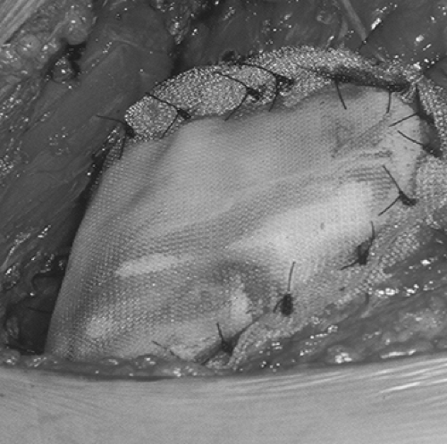 Figure 50-1. Methyl methacrylate (Marlex mesh) sandwich reconstruction after an anterior sternal and chest wall resection for an angiosarcoma related to radiation therapy for breast cancer. |
Historically, bone, diced cartilage, metal sheets, superstructures with autogenous rib graft, fascia lata, Teflon, and numerous other substances have been used with minimal success. With modern surgical technique, a wide range of reconstructive options are at the surgeon’s disposal; hence it is imperative that the appropriate procedure for a given patient be selected. In general, all full-thickness skeletal defects that have the potential for paradox should be reconstructed. The decision not to reconstruct the skeleton depends on the size and location of the defect. For small defects (<5 cm) or those located posteriorly under the scapula above the fourth rib (after resection of Pancoast tumors), the skeletal component can be ignored and the defect closed with only soft tissue. For patients with large chest wall defects (>5 cm), stabilization of the defect is indicated. Arnold and Pairolero5 state that most patients can tolerate sternectomy
or resection of four to six ribs at the cartilage level without experiencing flail chest or respiratory insufficiency postoperatively. Although the number of resected ribs is an important indicator for mesh usage, there does not appear to be a direct association between the number of ribs resected and the need for mesh reconstruction. This is likely because of the presence of additional factors that influence chest wall stability. Such factors include defect location and history of radiation. Location of the chest wall defect does appear to influence the need for skeletal stabilization, with mesh reconstruction being required more often for lateral defects. The lack of sternal or spinal stability in that location renders the patient more prone to flail chest deformities following chest wall resection.
or resection of four to six ribs at the cartilage level without experiencing flail chest or respiratory insufficiency postoperatively. Although the number of resected ribs is an important indicator for mesh usage, there does not appear to be a direct association between the number of ribs resected and the need for mesh reconstruction. This is likely because of the presence of additional factors that influence chest wall stability. Such factors include defect location and history of radiation. Location of the chest wall defect does appear to influence the need for skeletal stabilization, with mesh reconstruction being required more often for lateral defects. The lack of sternal or spinal stability in that location renders the patient more prone to flail chest deformities following chest wall resection.
Stay updated, free articles. Join our Telegram channel

Full access? Get Clinical Tree


