, John H. Stone2 and John H. Stone3
(1)
Harvard Medical School Division of Rheumatology, Department of Medicine, Massachusetts General Hospital, Boston, MA, USA
(2)
Harvard Medical School, Boston, USA
(3)
Clinical Rheumatology, Division of Rheumatology, Allergy and Immunology, Department of Medicine, Massachusetts General Hospital, Boston, MA, USA
Abstract
Cardiac manifestations of rheumatic diseases are varied and can involve every aspect of the cardiovascular system. Although the diseases can affect any organ system the majority of patients have joint pain or constitutional symptoms as part of their presentation. The rheumatologic diseases are important to recognize as treatment of the underlying disease can alleviate its cardiovascular manifestations.
Abbreviations
ANA
Antinuclear antibody
ANCA
Anti-neutrophil cytoplasmic antibodies
anti-CCP
Anti-cyclic citrullinated peptide antibody
aPL
Antiphospholipid antibodies
AV
Atrioventricular
bpm
Beats per minute
CAD
Coronary artery disease
CNS
Central nervous system
CSS
Churg Strauss Syndrome
CVD
Cardiovascular disease
GI
Gastrointestinal
HLA
Human leukocyte antigen
ILD
Interstitial lung disease
IVIG
Intravenous immune globulin
MI
Myocardial infarction
MRI
Magnetic resonance imaging
NSAID
Non-steroidal anti-inflammatory drug
RA
Rheumatoid arthritis
RF
Rheumatoid factor
SLE
Systemic Lupus Erythematosus
TNF
Tumor necrosis alpha
Introduction
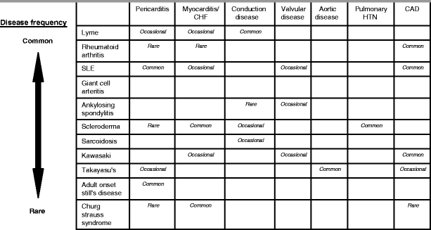
Cardiac manifestations of rheumatic diseases are varied and can involve every aspect of the cardiovascular system (Table 31-1). Although the diseases can affect any organ system, the majority of patients have joint pain or constitutional symptoms as part of their presentation. The rheumatologic diseases are important to recognize as treatment of the underlying disease can alleviate its cardiovascular manifestations.

Table 31-1
Cardiac involvement in rheumatic diseases

Lyme Disease
A.
Clinical presentation
Early localized disease – erythema migrans rash, arthralgias (without joint effusions) and constitutional symptoms.
Early disseminated disease – cranial neuropathies, meningitis and peripheral neuropathy.
Late disseminated disease – large joint arthritis (with effusions), encephalopathy, peripheral neuropathy.
B.
Cardiac manifestations – occur in early disseminated disease (1–2 months after the onset of symptoms) in 1.5–10 % of untreated patients [1].
Atrioventricular (AV) block is most common, present in 20–50 % of patients, with up to 30 % of symptomatic patients requiring temporary pacing.
Myopericarditis may be present but tends to be relatively mild with <15 % of patients demonstrating signs of heart failure and only mild left ventricular systolic dysfunction.
C.
Diagnosis
Lyme antibody confirmed by Western blot.
D.
Treatment
Mild manifestations (First degree AV block) – Doxycycline for 4 weeks.
Severe manifestations (Second or Third degree AV block) – Ceftriaxone for 4 weeks.
Rheumatoid Arthritis (RA)
A.
Clinical presentation
Women > men, incidence highest 50–70 years old.
Articular manifestations – symmetric, polyarticular arthritis.
Extra-articular manifestations – nodules (skin and lung), pleuritis, interstitial lung disease (ILD), scleritis.
B.
Cardiac manifestations
Coronary artery disease (CAD) – patients with RA have increased incidence of MI and mortality from cardiovascular disease [2].
Pericarditis and myocarditis – occur in <1 % of patients, usually in those with seropositive, erosive disease.
C.
Diagnosis
Rheumatoid factor (RF; less specific) or anti-cyclic citrullinated peptide antibody (anti-CCP; more specific). Combined sensitivity ∼80 %.
Joint erosions on radiographs.
D.
Treatment
CAD – treatment of RA may improve cardiovascular outcomes [3].
Pericarditis/myocarditis – prednisone 1 mg/kg ± steroid sparing immunomodulator.
Systemic Lupus Erythematosus (SLE)
A.
Clinical Presentation
Women > men, incidence highest 30–50 years old
Skin disease – malar rash, photosensitivity, discoid rash
Oral ulcers
Arthritis – two or more swollen and tender joints
Serositis – pleuritis or pericarditis
Renal involvement – proteinuria
Neurologic involvement – seizures or psychosis
Hematologic – leucopenia, thrombocytopenia or hemolytic anemia
Antiphospholipid syndrome – arterial or venous thrombosis, thrombocytopenia, second-trimester pregnancy losses
B.
Cardiac manifestations
CAD – patients at increased risk for myocardial infarction (MI) and cardiovascular mortality independent of traditional risk factors [4].
Pericarditis – most common cardiac manifestation occurring in 20–50 % of patients. Tamponade is rare (<10 %) [5].
Valvulitis – manifests as nodules or sterile vegetations (Libman-Sacks endocarditis) on aortic and mitral valves in 3–19 % of patients undergoing echocardiography [5]. Patients with high-titer antiphospholipid antibodies are at higher risk.
Myocarditis – < 10 % of patients [6]. Typically occurs with other disease manifestations. Rarely initial manifestation.
C.
Diagnosis
Clinical diagnosis. Most patients meet 4 of 11 classification criteria which include clinical manifestations (listed above) and the following:
Antinuclear antibody (ANA) positivity (>95 % of patients)
Positive antibodies – anti-Smith or double-stranded DNA antibody or positive tests for antiphospholipid antibodies (aPL).
Optimal testing for aPL includes:
Anticardiolipin antibodies
Lupus anticoagulant assay
Anti-beta-2-glycoprotein I antibodies
It is worth noting that the classification criteria are not diagnostic criteria, and some patients have SLE without fulfilling the classification criteria. Conversely, other patients fulfill classification criteria but do not have SLE.
D.
Treatment
Pericarditis – Non-steroidal anti-inflammatory drugs (NSAIDs) and low dose corticosteroids if NSAID refractory.
Valvulitis – anticoagulation if thrombotic events.
Myocarditis – high dose corticosteroids ± another immunosuppressive agent.
Giant Cell Arteritis
A.
Clinical manifestations
Occurs in individuals >50 years old.
Fever, headache, visual changes, jaw claudication, shoulder/hip girdle stiffness and pain.
B.
Cardiac manifestations
Aortitis – 18 %, thoracic > abdominal, 5 % with dissection [7].
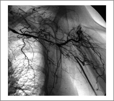
Large vessel vasculitis – 13 %, leading to symptomatic stenosis (most commonly arm claudication) [7] (Fig. 31-1).

Figure 31-1
Conventional angiogram from a patient with giant cell arteritis. There are several long, smooth stenoses of the left subclavian artery. Multiple collateral blood vessels are seen in the region of the shoulder. Such a patient will have greatly diminished or absent pulses in the arm but ample blood flow even to the distal extremity because of the collaterals. Re-vascularization attempts are usually not indicated, ineffective, and ill-advised
Coronary ischemia due to ostial left main and/or right coronary obstruction.
C.
Diagnosis
Temporal artery biopsy demonstrating granulomatous vasculitis.
Erythrocyte sedimentation rate elevated in 96 % [8].
D.
Treatment
Prednisone 1 mg/kg. ± Aspirin 81mg
Ankylosing Spondylitis
A.
Clinical presentation
Men > women, incidence highest 20–30 years old.
Axial symptoms – low back/buttock pain due to sacroiliitis (worse with rest).
Peripheral symptoms – asymmetric lower extremity oligoarthritis and enthesitis.
Extraarticular symptoms – uveitis, inflammatory bowel disease.
B.
Cardiac manifestations


Aortic regurgitation – 5–13 % of patients (typically mild) [5].
Conduction abnormalities – ECG abnormalities in up to 20 %, but symptomatic AV block rare [5].< div class='tao-gold-member'>Only gold members can continue reading. Log In or Register to continue
Stay updated, free articles. Join our Telegram channel

Full access? Get Clinical Tree


