and Jagmeet P. Singh2
(1)
Harvard Medical School Cardiology Division, Department of Medicine, Massachusetts General Hospital, Boston, MA, USA
(2)
Harvard Medical School Cardiac Arrhythmia Service, Cardiology Division, Department of Medicine, Massachusetts General Hospital, Boston, MA, USA
Abstract
Bradyarrhythmias are a heterogeneous group of cardiac rhythm disturbances which are implicated in over 40 % of sudden cardiac deaths in the hospital. Broadly classified, they are manifestations of either a failure of cardiac impulse generation or impulse propagation. They may be physiologic and benign, as in sinus bradycardia and sinus arrhythmia in athletes, or pathologic and warranting intervention, as in compromising bradycardia from sinus node dysfunction or ventricular asystole from high-grade atrioventricular (AV) block after an anterior myocardial infarction (MI).
The purpose of this chapter is to review the pathophysiology of various bradyarrhythmias and to review treatment options available. In addition, a brief overview of pacemakers and cardiac resynchronization therapy (CRT) will be provided.
Abbreviations
ACC
American College of Cardiology
AF
Atrial fibrillation
AHA
American Heart Association
AV
Atrioventricular
BPEG
British Pacing Electrophysiology Group
bpm
Beats per minute
CHB
Complete heart block
CL
Cycle length
CRT
Cardiac resynchronization therapy
cSNRT
Corrected sinoatrial node recovery time
ECG
Electrocardiogram
EP
Electrophysiologic
FDA
Food and Drug Administration
HCM
Hypertrophic cardiomyopathy
HF
Heart failure
HR
Heart rate
HRS
Heart Rhythm Society
ICD
Implantable cardioverter defibrillator
LAFB
Left anterior fascicular block
LBBB
Left bundle branch block
LPFB
Left posterior fascicular block
LQT1
Long QT syndrome type 1
LV
Left ventricular
LVEF
Left ventricular Ejection fraction
MI
Myocardial infarction
NASPE
North American Society for Pacing and Electrophysiology
NYHA
New York Heart Association
PPM
Permanent pacemakers
RA
Right atrium
RBBB
Right bundle branch block
RV
Right ventricle
SA
Sinoatrial
SACT
Sinoatrial conduction time
SND
Sinus node dysfunction
SNRT
Sinoatrial node recovery time
VT
Ventricular tachycardia
Introduction
Bradyarrhythmias are a heterogeneous group of cardiac rhythm disturbances which are implicated in over 40 % of sudden cardiac deaths in the hospital. Broadly classified, they are manifestations of either a failure of cardiac impulse generation or impulse propagation. They may be physiologic and benign, as in sinus bradycardia and sinus arrhythmia in athletes, or pathologic and warranting intervention, as in compromising bradycardia from sinus node dysfunction or ventricular asystole from high-grade atrioventricular (AV) block after an anterior myocardial infarction (MI).
The purpose of this chapter is to review the pathophysiology of various bradyarrhythmias and to review treatment options available. In addition, a brief overview of pacemakers and cardiac resynchronization therapy (CRT) will be provided.
Disorders of Impulse Generation
Sinus Arrhythmia
Definition: Phasic change in heart rate (HR) due to normal respiration
Pathophysiology: Thought to be due to reflex inhibition of vagal nerve tone during inspiration—leading to increase in HR during inspiration and slowing during respiration—thought to help improve and synchronize alveolar gas exchange [1]
Normal sinus arrhythmia
Most pronounced in the young
May be associated with sinus pauses for ≥ 2 s
Abolishment of sinus arrhythmia
Can be achieved through parasympathetic blockade by atropine
Autonomic denervation after cardiac transplant
Depression of respiratory sinus arrhythmia after MI is associated with an increased risk of sudden cardiac death [2]
Non-respiratory sinus arrhythmia [3]
In contrast to sinus arrhythmia, non-respiratory sinus arrhythmia is the change of p-p intervals varying at random
May reflect digitalis toxicity, intracranial hemorrhage, or ischemic heart disease
Treatment: respiratory sinus arrhythmia is usually not pathologic, even when associated with sinus pauses. When sinus arrhythmia coexists with symptomatic atrial tachyarrhythmias—as sometimes occurs in the case of young athletes—detraining with resultant de-conditioning may resolve the issue.
Sinus Bradycardia
Definition: Arbitrarily defined as sinus node impulse rate ≤ 60 beats per minute (bpm)
Pathophysiology: Sinus bradycardia or sinus pauses rarely cause hemodynamic instability, except when associated with extracardiac disturbance. For example:
Increased in vagal tone, includes:
Nausea and vomiting
Bowel obstruction
Urinary retention
Intracranial mass
Special case: Carotid sinus hypersensitivity
Sometimes considered a variant of vasovagal syncope, occurs more frequently in elderly patients and manifests as profound sinus bradycardia with sinus pauses from pressure on the carotid sinus
Dual-chamber pacing indicated for patients in whom recurrent syncope caused by spontaneously occurring carotid sinus stimulation and carotid sinus pressure induces ventricular asystole lasting ≥ 3 s present (Class I recommendation; see further details below) [4]
Sinus bradycardia can be exaggerated through parasympathomimetic or sympatholytic effect of drugs, notably:
β-blockers and calcium-channel blockers
Digoxin
Treatment: Identification of underlying etiology and avoidance of culprit agents is first-line treatment.
Minimally symptomatic sinus bradycardia with HR less than 40 bpm while awake is Class IIb indication for PPM [4].
Sinus Node Dysfunction (SND)
Definition: First described as sick sinus syndrome by Ferrer in 1968 [5], SND subsumes a constellation of abnormalities of the sinus node and surrounding atrial tissue characterized by sinus arrest, inappropriate sinus bradycardia (in the absence of drugs), and chronotropic incompetence. It is often coupled with the concurrent rise of subsidiary pacemakers leading to coexisting atrial tachyarrhythmias (hence, the term tachycardia-bradycardia syndrome).
Typically diagnosed in seventh and eight decades of life
Median annual incidence of complete AV block of 0.6 % and total prevalence of 2.1 %, suggestive of concurrent specialized conduction system degeneration [6]
Pathophysiology: Most commonly driven by senescence, SND may occur at any age due to destruction of sinus node cells through infiltration, collagen vascular disease, trauma, ischemia, infection or idiopathic degeneration [7]. Drugs can often exacerbate underlying SND (see Table 25-1).
Table 25-1
Cardioactive drugs that may induce or Worsen sinus node dysfunction
Β Blockers
Calcium channel blockers (e.g., verapamil, diltiazem)
Sympatholytic antihypertensives (e.g., α-methyldopa, clonidine, guanabenz, reserpine)
Cimetidine
Lithium
Phenothiazines (rarely)
Antihistamines
Antidepressants
Antiarrhythmic agents
May cause sinus node dysfunction (SND) in normal subjects: amiodarone
Frequently worsens mild SND: flecainide, propafenone, sotalol
Infrequently worsens mild SND: digitalis, quinidine, procainamide, disopyramide, moricizine
Rarely worsens mild SND: lidocaine, phenytoin, mexiletine, tocainide
Opiod blockers
Predominant clinical manifestations of SND include:
Frequent sinus pauses, sinus arrest, or sinus exit block
Inappropriate and severe sinus bradycardia with chronotropic incompetence
Episodes of bradycardia alternating with atrial tachyarrhythmias (usually atrial fibrillation (AF), although may be other supraventricular arrhythmias)
AF with a slow ventricular response or with very slow recovery after spontaneous conversion or cardioversion to revert to sinus rhythm
Diagnosis: Usually made based on elicitation of presyncopal symptoms or palpitations and confirmation on electrocardiogram (ECG). Other options include
Ambulatory ECG monitoring
Exercise testing to evaluate chronotropic competence
Electrophysiologic (EP) study may be diagnostic, and there is a class I indication to pursue EP study in patients with symptomatic bradycardia in whom a causal relationship between SND and symptoms has not been established [8]. Criteria evaluated include:
Sinoatrial node recovery time (SNRT): SA node is overdrive suppressed with atrial pacing, and the time from last paced atrial beat to the first spontaneous sinus beat is measured. Centers differ on normal SNRT, although <1,500 ms is conventional. A corrected SNRT (cSNRT) is the SNRT minus the sinus cycle length (CL), and is typically < 550 ms
Sinoatrial conduction time (SACT): is the time required for the sinus impulse to capture the atrium. Typically it is between 50 and 115 ms, and is often prolonged during SA block
Treatment: Largely depends on the diagnosis of symptomatic bradycardia, for which the only effective treatment is permanent cardiac pacing. Guideline recommendations are presented below [4]
Class I indications for permanent pacing in SND:
SND with documented symptomatic bradycardia, including frequent sinus pauses that produce symptoms
Symptomatic chronotropic incompetence
Symptomatic sinus bradycardia from required drug therapy for medical conditions
Class II indications for permanent pacing in SND:
SND with HR < 40 bpm, when symptoms are consistent with bradycardia, although the actual presence of bradycardia has not been documented
Syncope of unexplained origin with abnormal EP study (class IIa)
HR < 40 bpm while awake with minimal symptoms (class IIb)
Disorders of Impulse Propagation
Disorders of impulse propagation may occur at any point in the conduction system. Importantly, conduction block is distinct from the normal physiologic phenomenon of interference, in which a preceding impulse causes a period of refractoriness due to inactivation of ion channels.
Sinoatrial Exit Block
Definition: also called SA exit block, it manifests as sinus arrest of variable length on surface ECG. Prevalence is 1 % in otherwise normal subjects [9].
Pathophysiology: defect of impulse propagation within the SA node
First-degree SA exit block cannot be detected on surface ECG because sinus node depolarization is not inscribed separately from atrial depolarization (i.e., the p wave)
Second-degree SA exit block
Type 1: progressive prolongation of conduction block within the sinus node until complete exit block occurs (surface ECG demonstrates progressive shortening of p – p intervals before block)
Type 2: spontaneous block of sinus impulse leading to sinus pause which is an exact multiple of the preceding p – p interval
Third degree SA exit block: simply manifests as sinus arrest, usually with eventual appearance of subsidiary pacemaker (i.e., junctional escape rhythm)
Treatment: Sinoatrial exit block is usually treated in the context of SND, as indicated above
Atrioventricular (AV) Block
Definition: By convention, first-degree ‘block’ refers to impulses which are delayed, second-degree block refers to intermittent block of impulse conduction, and third-degree to complete block. Further specific terminology is described below.
First-degree AV block defined as PR interval > 0.20 s; generally felt due to block at the level of the AV node, although when associated with bundle branch block, may occur further down in the His-Purkinje system. Prevalence is 0.65 % in healthy adults [10]. Largely benign by itself, recent data from the Framingham cohort suggest that PR prolongation may be associated with increased risks AF, pacemaker implantation, and all-cause mortality over time [11]
Second-degree AV block was first classified into two types by Mobitz in 1924
Mobitz Type 1 (Wenkebach) AV block: characterized by progressive prolongation of the PR interval before non-conduction. Also generally associated with block at the level of the AV node.
Progressive shortening of R – R intervals prior to a dropped beat; shorter PR interval immediately after dropped beat
Irrespective of QRS width, usually represents an appropriate physiologic response to increasing HR through decremental conduction in the AV node
Mobitz Type 2: characterized by sudden non-conduction of atrial impulse without change in preceding PR interval. Usually represents infranodal disease.
Care should be taken to differentiate Mobitz II from a premature atrial complex (examine preceding p – p intervals) which causes physiologic interference and not conduction block
Some authors refer to multiple consecutive non-conducted impulses as ‘high-degree’ or ‘advanced’ heart block prior to true third-degree AV block
In the setting of AF, a prolonged pause ≥ 5 s is suggestive of underlying advanced second-degree AV block
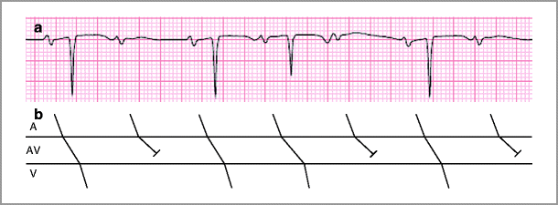
2:1 AV block: characterized by sudden non-conduction of atrial impulse without change in preceding PR interval after a single QRS complex. Based on surface ECG, it is difficult to discern whether location of block is within the AV node or below the level of the node (i.e., infrahisian). In patients with 2:1 AV block, evaluation of contemporaneous conduction disturbances (e.g., Wenkebach-type Mobitz 1) used to help infer level of block (see Fig. 25-1)

Figure 25-1
(a) Ladder diagram of 2:1 AV block and Wenkebach type block. (b) The rhythm strip shows a 2:1 block followed by short-stretch of Wenkebach and followed again with 2:1 block. The location of the block is inferred to be in the AV node due to the presence of Wenkebach, although cannot be determined conclusively without further information
Third-degree AV block, or complete heart block, occurs with absence of atrial impulse propagation to the ventricles and may manifest as ventricular standstill in the absence of an escape rhythm. When reversible etiologies are present (e.g., electrolyte disturbance, non-anterior ischemia), temporary pacing is usually indicated
Pathophysiology: There are numerous potential etiologies for AV block.
Physiologic AV block (first-degree of second-degree Type 1) is commonly due to enhanced vagal tone.
Idiopathic fibrosclerosis of the conduction system (i.e., Lev’s disease affecting the old and Lenegre’s affecting the young),
Infiltrative cardiomyopathy such as amyloidosis or sarcoidosis
Peri-AV nodal inflammation
Lyme disease
Myocarditis
Systemic lupus erythematosus
Dermatomyositis
Endocrinologic states
Thyroid storm or myxedema
Severe electrolyte disturbance
Hyperkalemia
Drug toxicity or overdose, particularly when agents are added in combination or if either renal or liver insufficiency occurs, which leads to accumulation of the drugs.
β blockers
Calcium channel blockers
Amiodarone
Digoxin
Iatrogenic etiologies of AV block are becoming increasingly common
Surgical or transcatheter aortic valve replacement
Alcohol septal ablation for hypertrophic cardiomyopathy
Transcatheter closure of ventricular septal defects
Complication of ablation during EP procedures.
Congenital etiologies are other rare but predictable causes of AV block:
Familial AV conduction block
Sequela of neonatal lupus syndrome (particularly in babies born of mothers that are positive for antinuclear antibodies SSA/Ro and SSB/La)
Hereditary neuromuscular diseases such as myotonic dystrophy
Myocardial ischemia is an important cause of AV and infranodal block
Many forms of AV block commonly seen in acute inferior MI, most often due to increased vagal tone, rarely due to AV nodal infarction
AV block and infranodal block due to acute anterior wall MI most often due to infarction of the conduction system
Treatment: the initial course of treatment is to identify and remove any potential reversible offending agents. The decision for permanent pacing is often left to the discretion of the cardiologist, based on an appreciation of the relative stability of the underlying rhythm and the risk associated with developing symptoms. Class I indications are as outlined in Table 25-2
Table 25-2
ACC/AHA/HRS Class I Recommendations for permanent pacing
Recommendations in acquired atrioventricular block in adults
Third-degree and advanced second-degree AV block at any anatomic level associated with bradycardia and symptoms (including heart failure) or ventricular arrhythmias presumed to be due to AV block
Third-degree and advanced second-degree AV block at any anatomic level associated with arrhythmias and other medical conditions that require drug therapy that results in symptomatic bradycardia
Third-degree and advanced second-degree AV block at any anatomic level in awake, symptom-free patients in sinus rhythm, with documented periods of asystole ≥ 3.0 s or any escape rate less than 40 bpm, or with an escape rhythm that is below the AV node
Third-degree and advanced second-degree AV block at any anatomic level in awake, symptom-free patients with AF and bradycardia with 1 or more pauses of at least 5 s or longer
Third-degree and advanced second-degree AV block at any anatomic level after catheter ablation of the AV junction
Third-degree and advanced second-degree AV block at any anatomic level with postoperative AV block that is not expected to resolve after cardiac surgery
Third-degree and advanced second-degree AV block at any anatomic level associated with neuromuscular diseases with AV block, such as myotonic muscular dystrophy, Kearns-Sayre syndrome, Erb dystrophy (limb-girdle muscular dystrophy), and peroneal muscular atrophy, with or without symptoms< div class='tao-gold-member'>Only gold members can continue reading. Log In or Register to continue
Stay updated, free articles. Join our Telegram channel

Full access? Get Clinical Tree

 Get Clinical Tree app for offline access
