Benign Tumors, Cysts, and Duplications of the Esophagus
Matthew A. Facktor
Mark R. Katlic
Benign esophageal tumors and cysts represent a collectively uncommon set of disease entities that do not necessarily require surgical resection. Most dedicated general thoracic surgeons will encounter these lesions at least a few times during their careers and may occasionally even be presented urgently with such a case after complications from endoscopic attempts at either biopsy or removal. The exact incidence of these benign esophageal lesions remains unknown owing to their rarity, inherently indolent course, and typically asymptomatic nature. Older autopsy studies report a general incidence of less than 1%, ranging anywhere between 0.006% and 0.59%,7,23 which is far less than that for esophageal carcinoma. Esophageal leiomyoma is by far the most common of these benign lesions, followed second by esophageal cysts and third by granular cell tumors. Generally the presence of symptoms serves as an indication for removal, and most authors agree that the mere presence of an esophageal cyst urges at least prophylactic resection to avoid potential complications.
This chapter describes the spectrum of benign esophageal tumors and cysts, their major diagnostic and distinguishing characteristics, the indications for intervention, and the currently employed techniques for removal. Perhaps the most dramatic advance in recent years related to this disease category is the diagnostic usefulness of endoscopic ultrasound (EUS), which, if performed and interpreted properly, may help avoid unnecessary operation in asymptomatic patients with benign esophageal leiomyomas. The other significant advance has been the continued expansion and application of minimally invasive approaches to removal, including thoracoscopy, laparoscopy, robotic-assisted surgery, and highly advanced endoscopic techniques. A suggested management algorithm is included at the end of the chapter to guide the reader through our recommendations in an organized manner.
Overview
Historically, Virchow first described the pathologic appearance of esophageal leiomyoma in 1867,7 Sauerbruch first reported the surgical resection of a benign esophageal tumor in 1932,7,29 and Ohsawa followed a year later, in 1933, with a similar report of surgical resection.29 Sixty years later, in 1992, the first two reports of minimally invasive thoracoscopic enucleation of esophageal leiomyoma were published.4,11 Collectively, benign esophageal tumors are uncommon and represent less than 1% of all clinically detected esophageal tumors.
Although few thoracic surgeons encounter a large number of these lesions, there are simple yet crucial details that, if known, will greatly help guide even the least experienced surgeon through diagnosis and management. First, and most important, symptomatic lesions (dysphagia, odynophagia, chest pain, obstruction, or bleeding) should generally be removed, pre- ferentially using minimally invasive techniques. Second, all cysts should generally be removed, regardless of the presence or absence of symptoms, in order to prevent potential future infection. Third, the most effective diagnostic tool is endoscopic ultrasound. Fourth, transthoracic needle aspiration should be avoided if at all possible unless the endosonographic appearance of the lesion is suspicious for malignancy. Finally, surgical removal, when necessary, involves adequate esophageal exposure, myomectomy of the muscularis propria, enucleation of the lesion with careful avoidance of mucosal injury, and optional closure of the incised muscle.
There are numerous types of benign esophageal lesions, both solid and cystic, and previous authors have described various classification schemes.7,24 Table 157-1 represents a typical list of the various subtypes. More important than memorizing lists, however, is for the surgeon to simply understand the normal layers of the esophageal wall, the means by which these tumors can be visualized nonsurgically, the potential symptoms they may cause, and the distinction between these benign lesions and their malignant counterparts. Potential malignant transformation of benign lesions has frequently been cited as an indication for resection, but it is important to realize that this has only rarely been reported, and malignant lesions other than squamous cell tumors or adenocarcinomas of the esophagus are themselves exceedingly rare. Expert endosonography should provide sufficient information to rule out obvious malignant invasion, regional lymphadenopathy, or suspicious heterogeneity of the tumor itself, all of which would lead to further evaluation of potential malignancy by obtaining tissue for pathologic diagnosis.
Table 157-1 Benign Esophageal Tumor Subtypes by Location Within the Esophageal Wall | |||||||||||||||
|---|---|---|---|---|---|---|---|---|---|---|---|---|---|---|---|
|
Imaging and Endoscopy
Benign esophageal lesions are frequently discovered incidentally on either radiographic or endoscopic examination; therefore their role in this disease process deserves a brief review. Imaging modalities to visualize these disease processes include barium esophagography, computed tomography (CT), esophagoscopy (EGD), and esophageal ultrasound (EUS). Detailed information regarding esophageal wall anatomy, imaging studies, and endoscopy procedures in general terms is covered elsewhere in this textbook. Of paramount importance, however, is the fact that the surgeon must be intimately familiar with anatomy of the esophageal wall in order to appropriately order, interpret, or personally perform these studies.
The three distinct layers of the esophageal wall include the mucosa, submucosa, and muscularis propria. The mucosa includes epithelium, basement membrane, lamina propria, and muscularis mucosa. The submucosa includes elastic fibers, collagen, and glands. The muscularis propria includes both inner circular and outer longitudinal layers, which transition from purely striated fibers in the cervical esophagus to purely smooth muscle fibers in the mid- to lower esophagus. The esophageal wall has no adventitial layer, so the paraesophageal tissue lies directly against the outer muscular wall.
Barium esophagography provides gross anatomic detail of the esophageal mucosa and is best performed as a biphasic examination, including both single- and double-contrast phases.21 Chest CT scanning is useful to demonstrate these lesions with their anatomic relationships, including some information regarding solid versus cystic characterization. CT cannot distinguish the layers of the esophageal wall, however, and cannot reliably distinguish thick cystic fluid from a solid tumor. Additionally, lesions arising from the midesophagus can be misread on CT scans as subcarinal lymphadenopathy.
EGD provides excellent direct visualization of the esophageal mucosa and a means by which superficial lesions may be biopsied or even removed entirely with advanced techniques. Endoscopic ultrasound, in addition to standard esophagoscopy, is capable of providing visualization of all layers of the esophageal wall, including paraesophageal lymph nodes outside the esophageal wall, and of providing guidance for direct needle aspiration of abnormalities when malignancy is suspected. Ultrasound is generally accepted in the radiologic community as more reliable than CT in determining whether a lesion is solid versus cystic, but the viscous nature of typical esophageal cyst contents may make this distinction difficult, despite EUS examination.
The distinct advantage of EUS is the ability to visualize each layer of the esophageal wall, which provides very important diagnostic information, as discussed later in this chapter. The layers are seen as alternating white (hyperechoic) and black (hypo- echoic) rings.20,28 The innermost ring is white and represents the superficial mucosa (epithelium and basement membrane). The second ring is black and represents the deeper mucosa (lamina propria and muscularis mucosa). The third ring is white and represents the submucosa. The fourth ring is black and represents the muscularis propria. The fifth and outermost ring is white and represents the paraesophageal tissue, which includes the paraesophageal lymph nodes.
Important additional information gleaned from EUS includes the general ultrasound characteristics of the lesion. Typically, homogenous lesions are benign, while heterogeneous lesions may be malignant. Benign lesions may demonstrate hypoechoic, hyperechoic, or indeterminate echogenicity.28 Most authors agree that heterogeneity and size >3 cm significantly raise the suspicion for malignancy, and therefore warrant attempts at fine-needle aspiration (FNA) for tissue diagnosis.20,28
Transesophageal needle biopsy is not generally favored in benign-appearing lesions for several important reasons. First, there is an inherent risk of infection due to the transmucosal nature of the procedure. Second, the inflammatory adhesions that naturally occur between the mucosa and the lesion as a result of instrumentation may complicate subsequent surgical resection by increasing the risk of mucosal injury during enucleation. Finally and perhaps most importantly, if the lesion appears both solid and benign to an experienced endosonographer, observation alone is warranted owing to the naturally indolent course of these tumors. More specific EUS tumor characteristics are discussed further in the subsequent sections.
Benign Tumors of the Mucosa
Benign tumors of the esophageal mucosa, because of the simple nature of their location, are typically visualized directly during esophagoscopy. These tumors are therefore amenable to biopsy with endoscopic pinch forceps for pathologic examination. Further examination with EUS may provide important additional diagnostic information.
Granular Cell Tumors
Granular cell tumors represent the third most common benign esophageal tumor (behind leiomyoma and cysts). These small, yellow, firm tumors have been found in numerous locations, including the gastrointestinal tract, respiratory tract, skin, and breast. Within the gastrointestinal tract, the distal third of the esophagus is the most common location, and they have been shown immunohistochemically to arise from a neural (Schwann cell) origin. Granular cell tumors are very uncommon, typically asymptomatic, small, and usually found incidentally either at EGD (Fig. 157-1) or autopsy. Infrequently, two or more lesions
may be identified in a single patient.6 Direct forceps biopsy may well be diagnostic, although not in all cases. Evaluation by EUS demonstrates a smooth lesion with origin from the inner two layers (less than 5% arise from the submucosa), and there may be an inhomogeneous, hypoechoic pattern.28 Atypical EUS features may suggest malignancy, which is very rare but generally confirmed pathologically by mitotic activity, large nuclear size, and nuclear pleomorphism.12 Clinical observation is recommended in the absence of symptoms, tumor size >1 cm, or atypical EUS features.15
may be identified in a single patient.6 Direct forceps biopsy may well be diagnostic, although not in all cases. Evaluation by EUS demonstrates a smooth lesion with origin from the inner two layers (less than 5% arise from the submucosa), and there may be an inhomogeneous, hypoechoic pattern.28 Atypical EUS features may suggest malignancy, which is very rare but generally confirmed pathologically by mitotic activity, large nuclear size, and nuclear pleomorphism.12 Clinical observation is recommended in the absence of symptoms, tumor size >1 cm, or atypical EUS features.15
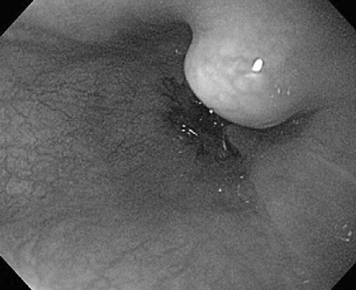 Figure 157-1. Typical endoscopic appearance of a granular cell tumor. (Photo courtesy of Dr. David Diehl, Geisinger Medical Center, Danville, PA.) |
Fibrovascular Polyps
Benign esophageal mucosal polyps arise at or near the cricoid cartilage in the cervical esophagus with extension into the lumen. The lesions are cylindrical and based on a long pedicle, with a histologic appearance that displays variable components, including fibrous, vascular, adipose, and neural tissue.22 Large tumors tend to cause dysphagia and/or obstruction of the esophageal lumen or rarely even regurgitation into the mouth with potential asphyxiation and sudden death.1,8 The intraluminal nature of these lesions makes them readily visible on a typical barium esophagram and even chest and/or neck CTs. EGD and EUS are not particularly helpful. Removal is generally recommended, especially if such lesions are detected at a large size, to prevent complications from obstruction or ulceration of the overlying mucosa.
Squamous Papillomas
Esophageal papillomas are rare, benign, sessile lesions of varying size, typically located in the distal esophagus. Although controversial, the lesions may arise as a result of infection with human papillomavirus,26,27,34 but chronic inflammation from sources such as gastroesophageal reflux disease has also been implicated.28 Endoscopic biopsy is critical to distinguish these lesions from superficial squamous cell carcinoma, and EUS may help rule out any invasive characteristics. Excision by either endoscopic or surgical techniques is recommended for symptomatic or histologically atypical lesions; otherwise the asymptomatic lesion may be followed clinically.
Stay updated, free articles. Join our Telegram channel

Full access? Get Clinical Tree


