Bedside Assessment of the Patient


Bedside Assessment of the Patient


Richard H. Kallet


Progress in the field of respiratory care has placed increasing demands on respiratory therapists (RTs) to develop competent bedside assessment skills. Decisions regarding when to initiate, change, or discontinue therapy depend on accurate clinical assessment. Although the physician has the ultimate responsibility for these decisions, RTs often participate in the clinical decision-making process. To fulfill this role effectively, the RT must assume responsibility for gathering and interpreting relevant bedside patient data.


Bedside assessment is the process of interviewing and examining a patient for signs and symptoms of disease and the effects of treatment. It is a cost-effective way of obtaining pertinent information about the patient’s health status. In many cases, bedside assessment provides the initial evidence that something is wrong and often helps establish the severity of the problem. In contrast to some diagnostic tests, bedside assessment techniques are of little risk to the patient.


Two key sources of patient data are the medical history and the physical examination. Data gathered initially by interview and physical examination help identify the need for subsequent diagnostic tests. After a tentative diagnosis is made, these assessment procedures also help the clinician to select the best approach to therapy. After a treatment regimen begins, these assessment procedures are repeated to monitor patient progress toward predefined goals.


The patient initially is assessed to identify the correct diagnosis. This initial assessment is most often performed by a physician. Exceptions may occur in emergency situations in which a physician is unavailable. In such cases, other health care personnel, such as nurses and RTs, may need to evaluate the patient rapidly to implement appropriate lifesaving treatment (e.g., cardiopulmonary resuscitation). After a tentative diagnosis is reached and the physician orders specific treatment, subsequent evaluations are made by health care personnel to monitor the patient during the hospital stay and to evaluate treatment results.


The skills of bedside assessment described here are not difficult to learn; however, mastery requires practice. Initially, students should practice the skills on healthy individuals. This practice helps improve technique and provides an understanding of normal variations. The ability to discriminate abnormal from the range of possible normal findings is an important skill that requires experience to master.


Interviewing the Patient and Taking a Medical History


Interviewing furnishes unique information because it provides the patient’s perspective. It serves the following three related purposes:


For these reasons, interviewing is a crucial aspect of general patient assessment.


Principles of Interviewing


Interviewing is a way of “connecting” with the patient. This connection is especially important for the patient who is under the stress of an illness because meaningful human contact lessens the patient’s sense of isolation. The factors that affect communication between the RT and the patient include the following:


Because of the above-listed factors, no two interviews are the same.


Although developing interviewing skills takes time and experience, beginners can get a head start by following a few basic guidelines and by becoming knowledgeable about the causes and characteristics of common cardiopulmonary symptoms. The following discussion provides some of the guidelines for interviewing and discusses common symptoms associated with diseases of the chest.


Structure and Technique for Interviewing


The ideal interview is one in which the patient feels secure and free to talk about important personal matters. Each interview should begin with the RT introducing himself or herself to the patient and stating the purpose of the visit. The introduction is done in the social space, approximately 4 to 12 feet from the patient. It begins the process of establishing a rapport with the patient and helps the patient feel more comfortable about answering personal questions. Pulling the curtain between the beds of a semiprivate room also may be helpful in making the patient feel more at ease with the interview (Box 15-1).


Box 15-1   Guidelines for Effective Patient Interviewing


Show Your Respect for the Patient’s Beliefs, Attitudes, and Rights


• Ensure the patient is appropriately covered.


• Position yourself so that eye contact is comfortable for the patient. (Ideally, patients should be sitting up, with their eye level at or slightly above yours.)


• Avoid standing at the foot of the bed or with your hand on the door because this may send the nonverbal message that you do not have time for the patient.


• Ask the patient’s permission before moving any personal items or making adjustments in the room.


• Remember that the patient’s dialog with you and his or her medical record are confidential. Share this information only with other health care providers who need to know about it, and do not share the information in a place where others can overhear the conversation.


• Be honest; never guess at an answer or information that you do not know; do not provide information beyond your scope of practice; providing new information to the patient is the privilege and responsibility of the attending physician.


• Make no moral judgments about the patient; set your values for patient care according to the patient’s values, beliefs, and priorities.


• Expect the patient to have an emotional response to illness and the health care environment.


• Listen, and then clarify and teach, but never argue.


• Adjust the time, length, and content of the interview to the patient’s needs.


Next, the RT moves into the personal space (2 to 4 feet from the patient) to begin the interview. In this space, the patient does not have to speak loudly in response to questions. The RT should assume a physical position at the same level with the patient (e.g., by sitting in a chair) before beginning the formal interview. Standing over the patient should be avoided because this position makes the patient feel inferior. Appropriate eye contact with the patient is essential for a quality interview. Eye contact gives the patient more confidence in the interviewer. Eye contact also allows the interviewer to see confusion, anger, frustration, and other emotions that may be expressed by the patient in response to questions.


Using neutral questions and avoiding leading questions during the interview is important. Asking the patient, “Is your breathing better now?” leads the patient toward a desired response and may elicit false information. Asking the patient, “How is your breathing now?” is a better way to get accurate information about the patient’s breathing (Box 15-2).


Box 15-2   Types of Questions Used in Patient Interviews


• Open-ended questions encourage patients to describe events and priorities as they see them, helping to bring out concerns and attitudes and to promote understanding. Questions such as “What brought you to the hospital?” or “What happened next?” encourage conversational flow and rapport, while giving patients enough direction to know where to start.


• Closed questions, such as “When did your cough start?” or “How long did the pain last?” focus on specific information and provide clarification.


• Direct questions can be open-ended or closed and always end in a question mark. Although they are used to obtain specific information, a series of direct questions or frequent use of the question “Why?” can be intimidating and cause the patient to minimize his or her responses to questions.


• Indirect questions are less threatening than direct questions because they sound like statements (e.g., “I gather your doctor told you to take the treatments every 4 hours”). Inquiries of this type also work well to confront discrepancies in the patient’s statements (e.g., “If I understood you correctly, it is harder for you to breathe now than it was before your treatment”).


• Neutral questions and statements are preferred for all interactions with the patient. “What happened next?” and “Can you tell me more about …?” are neutral, open-ended questions. A neutral, closed question may give the patient a choice of responses, while focusing on the type of information desired (e.g., “Would you say there was a teaspoon, a tablespoon, or a half cup?”). Leading questions, such as “You didn’t cough up blood, did you?” should be avoided because they imply an answer.


Common characteristics of symptoms can be identified by asking questions such as the following during the interview:


Identifying these characteristics of any new symptom can be helpful in recognizing the cause and potential therapy; this is primarily the role of the attending physician but sometimes falls to other clinicians in certain settings. Once the symptom or symptoms are established and therapy is started, other questions are used to evaluate the changes in the symptoms over the course of the hospital stay. For example, the clinician may ask, “Has the symptom changed in any way since admission?” or, “Does the therapy seem to make a difference?”


The best interview techniques are of no value if the interviewer is not knowledgeable about the pathophysiology and characteristics of common cardiopulmonary symptoms. The interview is a series of focused questions that pursue specific information related to a tentative diagnosis. The ability to ask the key questions at the right time comes from experience and familiarity with the signs and symptoms of lung disease.


Common Cardiopulmonary Symptoms


Dyspnea


Dyspnea is a general term describing the sensation of breathing discomfort. It is the most important symptom that the RT is called on to assess and treat. Dyspnea is a subjective experience and should not be inferred from observing the patient’s breathing pattern. Analogies often have been made between dyspnea and pain. Both sensations possess qualitatively distinct features and varying intensity. Similar to pain, dyspnea causes suffering. As breathing is the primordial sensation of life, dyspnea often is perceived as life-threatening and may provoke a profound sense of dread.


The term dyspnea also is used specifically to describe difficulty in the mechanical act of breathing. The simplest explanation is that the effort to breathe is proportionally greater than the tidal volume achieved. A person’s perception of breathing is a complex balance between the following three factors:


When the neuronal signals governing these sensations become unbalanced, breathing is perceived to be abnormal and unpleasant. The technical name for this imbalance is neuromechanical dissociation. Individuals normally experience this form of dyspnea only in unusual circumstances, such as when trying to breathe through a straw or when wearing a restrictive garment.


Breathlessness

Breathlessness is the specific sensation of an unpleasant urge to breathe. It is believed to be the conscious perception of intense neural discharge to the respiratory muscles. Breathlessness can be triggered by acute hypercapnia and acidosis and by hypoxemia. A normal experience of breathlessness is the unpleasant throbbing sensation that accompanies prolonged breath holding or feeling “winded” during strenuous physical exercise. However, it is not known how closely normal encounters with breathlessness resemble the sensation that arises during disease because dyspnea in patients with cardiopulmonary disease also is affected by other stimuli that contribute to both the quality and the intensity of the sensation. These include stimuli arising from hypoxemia, irritant receptors in the lungs and airways, and receptors in the blood vessels and heart.


Ultimately, dyspnea and breathlessness are magnified by the emotional distress that accompanies them. This distress is influenced by situation, knowledge, and control. In other words, a healthy person can quickly identify the source of breathlessness and arrest the symptom simply by stopping exercise or the breath hold. In contrast, a patient with cardiopulmonary disease may be unable to control the symptom, let alone be able to identify the source. These factors have a profound emotional impact that must be appreciated by the RT.


Positional Dyspnea

Dyspnea may be present only when the patient assumes the reclining position, in which case it is referred to as orthopnea. Orthopnea is common in patients with congestive heart failure (CHF); it apparently is caused by the sudden increase in venous return that occurs with reclining. The failing left ventricle is unable to accommodate the increased venous return, resulting in pulmonary vascular congestion and dyspnea. Orthopnea is also a symptom of bilateral diaphragmatic paralysis.


Dyspnea in the upright position is known as platypnea. This unusual symptom may accompany arteriovenous malformations in the lung, such as occur in chronic liver disease (hepatopulmonary syndrome), and some hereditary conditions. Platypnea may be accompanied by orthodeoxia, which is oxygen desaturation on assuming an upright position.


Language of Dyspnea

Dyspnea is a subjective experience, and patients possess a nuanced language to describe their sensations. RTs should ask specific questions about the quality and characteristics of the patient’s dyspnea. In this way, the RT might gain insight into the mechanism provoking dyspnea. As the patient describes the sensations, the RT should try to categorize each according to a particular aspect of breathing such as inspiration, expiration, respiratory drive, or lung volume. A remark such as, “I feel that my breath stops,” reflects a problem with inspiration, whereas the remark, “my breath does not go all the way out,” suggests a problem with expiration. Statements such as, “I can’t catch my breath,” suggest that respiratory drive is elevated (i.e., breathlessness).


Different types of lung diseases often evoke unique sensations that may provide clues about the underlying pathophysiology. Patients with asthma frequently complain of chest tightness. In contrast, patients with interstitial lung disease tend to focus on the sensations of increased work of breathing, shallow breathing, and gasping. Patients with CHF are seemingly unique in frequently feeling suffocated. Although the language used to describe dyspnea provides helpful clues, the RT should keep in mind that many lung diseases evoke common sensations.


Patients with cardiopulmonary disease frequently experience several unpleasant breathing sensations simultaneously. A particular sensation may be more prominent than others and may change over time. As mentioned previously, patients with asthma typically complain first about the sensation of chest tightness. However, as bronchoconstriction worsens and the lungs become more hyperinflated, patients often begin to focus more on the sensation of excessive work of breathing, air hunger, and the inability to take a deep breath.


Assessing Dyspnea in the Interview

The assessment of dyspnea is largely determined by the situation. When conducting an interview, the RT should pay particular attention to whether the patient can speak in full sentences. It may be very difficult for patients with severe dyspnea from any cause to speak more than a few words at a time. In this situation, the initial interview should be curtailed, and treatment should be initiated as soon as possible. Questions should be brief and limited to the quality and intensity of dyspnea and the circumstances of symptom onset. Also, the assessment of dyspnea should occur simultaneously with a gross examination of the patient’s breathing pattern (see later section of this chapter). Assessment of dyspnea that arises acutely in patients without a prior history of cardiopulmonary disease typically does not require the same detail as assessment in patients with long-standing cardiopulmonary or neurologic disease.


In patients with chronic cardiopulmonary disease, a detailed and systematic history should cover four major areas, as follows:


1. The RT should ask what activities of daily living tend to trigger episodes of dyspnea. For example, is dyspnea triggered by walking on flat surfaces, by climbing stairs, by bathing, by dressing?


2. The RT should ask how much exertion is required for the patient to stop to catch his or her breath with different activities. Does the patient need to stop after walking up one flight of stairs or one step? Dyspnea provoked by less strenuous activities indicates more advanced disease.


3. The RT should ask whether the quality or the sensation of breathing discomfort varies with different activities.


4. To gain a better understanding of the patient’s history, the RT should ask the patient to recall when dyspnea first began and how it has evolved over time. Has dyspnea progressed slowly or rapidly? How long has this progression taken place: over a period of months or years? Has there been a dramatic change in the intensity of dyspnea over recent months, weeks, days, or even within the past few hours?


Beyond the information gleaned, a detailed conversation about a patient’s struggle with dyspnea allows the patient to share his or her experience and decreases the patient’s sense of isolation.


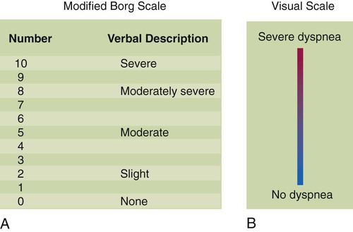
The intensity of dyspnea can be documented using a numeric intensity or visual analog scale (Figure 15-1). Such scales provide a way to evaluate the patient’s response to treatment over time. These scales are important because objective lung function measurements (e.g., pulmonary function tests, PaO2) seldom correlate with the degree of dyspnea in many patients.


Psychogenic Dyspnea: Panic Disorders and Hyperventilation

There are perplexing situations in which patients with normal cardiopulmonary function complain of intense dyspnea or suffocation. This condition is known as psychogenic hyperventilation syndrome and is associated with panic disorders. Hyperventilation may coincide with other symptoms such as chest pain, anxiety, palpitations and paresthesia (the sensation of tingling and numbness in the extremities that often accompanies respiratory alkalosis). This syndrome may be either sporadic or chronic and often is self-perpetuating.


Anxiety often is accompanied by breathlessness and hyperventilation. The resulting respiratory alkalosis amplifies the sensation of breathlessness and provokes more anxiety, increasing the intensity of hyperventilation. The classic homespun remedy of having the patient slowly rebreathe into a paper bag holds merit because this can arrest the respiratory alkalosis and help break the cycle. However, rebreathing techniques may require formal behavioral therapy and may not be appropriate in the hospital setting. This condition usually is treated clinically by administering judicious amounts of anxiolytic agents.


The RT always must approach any situation involving hyperventilation or dyspnea as if it had a pathophysiologic basis. The first priority is to measure the vital signs, including arterial oxygen saturation, and perhaps a 12-lead electrocardiogram and arterial blood gases. A psychogenic source should be considered only after a pathogenic source for hyperventilation or dyspnea has been ruled out. Intense pain or fear may provoke anxiety and hyperventilation. The RT must work in concert with nursing and physician colleagues to determine the root cause of any hyperventilation syndrome.


Cough


A cough is the most common, yet nonspecific symptom seen in patients with pulmonary disease. Coughing is a forceful expiratory maneuver that expels mucus and foreign material from the airways. It usually occurs when the cough receptors are stimulated by inflammation, mucus, foreign materials, or noxious gases. The cough receptors are located primarily in the larynx, trachea, and larger bronchi.


The effectiveness of a cough depends on the ability of the individual to take a deep breath, lung elastic recoil, expiratory muscle strength, and level of airway resistance. The ability to take a deep breath or exhale forcefully is often impaired in patients with neuromuscular disease. An effective cough also is impaired secondary to pain; this is typically seen in the early postoperative period in patients following upper abdominal surgery or thoracic surgery or after trauma. Often expiratory flow is limited by factors such as bronchospasm (e.g., asthma) and reduced lung elastic recoil (as in emphysema). Patients with an inadequate ability to cough because of impairment of these factors often have problems with retained secretions and are more prone to the development of pneumonia.


Important characteristics of the patient’s cough to identify include whether it is dry or loose, productive or nonproductive, and acute or chronic and whether it occurs more frequently at particular times (i.e., day or night). Knowledge of such details may help in determining the cause of the cough. A dry, nonproductive cough is typical for restrictive lung diseases such as CHF or pulmonary fibrosis. A loose, productive cough is more often associated with inflammatory obstructive diseases such as bronchitis and asthma. The most common cause of an acute, self-limited cough is a viral infection of the upper airway. Common causes of chronic coughing include asthma, postnasal drip, chronic bronchitis, and gastroesophageal reflux,1 although combinations of these often exist.2 Cough is also associated with the use of certain medications for hypertension (e.g., angiotensin-converting enzyme inhibitors).3


Sputum Production


Healthy airways produce mucus daily. Normally, the quantity of this mucus is minimal, and it is not enough to stimulate the cough receptors. Mucus is gradually moved to the hypopharynx by the mucociliary escalator, where it is either swallowed or expectorated. Disease of the airways may cause the mucous glands, which line the airways, to produce an abnormally increased amount of mucus, which usually stimulates the cough receptors and causes the patient to generate a loose, productive cough. This cough is seen in acute bronchitis or asthma attacks brought on by airway infection.


RTs need to be aware of the terminology associated with sputum. Technically, mucus from the tracheobronchial tree that has not been contaminated by oral secretions is called phlegm. Mucus that comes from the lung but passes through the mouth as it is expectorated is sputum. Because this is how most mucus samples from the lung are obtained, the term sputum is used in this chapter. Sputum that contains pus cells is said to be purulent, suggesting a bacterial infection. Purulent sputum appears thick, colored, and sticky. Sputum that is foul-smelling is said to be fetid. Sputum that is clear and thick is mucoid and is commonly seen in patients with airways disease (i.e., asthma). Changes in the color, viscosity, or quantity of sputum produced are often signs of infection and must be documented and reported to the physician.


Hemoptysis


Coughing up blood or blood-streaked sputum from the lungs is referred to as hemoptysis. Blood-streaked sputum is common in patients with pulmonary disease. Frank hemoptysis is the presence primarily of blood in the expectorant. Hemoptysis is characterized as massive when more than 300 ml of blood is expectorated over 24 hours, and this represents a medical emergency. Hemoptysis must be distinguished from hematemesis, which is vomiting blood from the gastrointestinal tract. Blood from the lung is often seen in patients with a history of pulmonary disease and may be mixed with sputum. Blood from the stomach may be mixed with food particles and occurs most often in patients with a history of gastrointestinal disease.


Nonmassive hemoptysis is caused most often by infection of the airways but also is seen in lung cancer, tuberculosis, blunt or penetrating chest trauma, and pulmonary embolism. Hemoptysis associated with infection usually is seen as blood-streaked, purulent sputum. Hemoptysis commonly is found in patients with bacterial pneumonia. Hemoptysis from bronchogenic carcinoma often is chronic and may be associated with a monophonic wheeze and cough. Common causes of massive hemoptysis include bronchiectasis, lung abscess, and acute or old tuberculosis.


Chest Pain


Most chest pain can be categorized as either pleuritic or nonpleuritic. Pleuritic chest pain usually is located laterally or posteriorly. It worsens when the patient takes a deep breath, and it is described as a sharp, stabbing type of pain. It is associated with diseases of the chest that cause the pleural lining of the lung to become inflamed, such as pneumonia or pulmonary embolism.


Nonpleuritic chest pain is located typically in the center of the anterior chest and may radiate to the shoulder or back. It is not affected by breathing, and it is described as a dull ache or pressure type of pain. A common cause of nonpleuritic chest pain is angina, which classically is a pressure sensation with exertion or stress and results from coronary artery occlusion. Other common causes of nonpleuritic chest pain include gastroesophageal reflux, esophageal spasm, chest wall pain (e.g., costochondritis), and gallbladder disease.


Mini Clini


Sudden Onset of Chest Pain


image Problem


The RT is called to the emergency department to see a 47-year-old man who came to the hospital with anxiety and chest pain. He is certain he is having a heart attack and demands immediate treatment. The attending physician is on the way to the hospital but has asked the nurse to call the RT in the interim. The RT places the patient on oxygen per protocol and asks him for details about the chest pain. The patient states that the pain is located laterally on the left side and increases with each inspiratory effort. The pain is sharp in nature. The patient’s vital signs are normal (including respiratory rate) except for a slight increase in heart rate. What is the most likely cause of this patient’s chest pain, given its characteristics? What should be done until the physician arrives?


Solution


Chest pain is a worrisome symptom because it can indicate a life-threatening problem or a less serious problem. Among the most serious problems are acute heart attack, pulmonary embolism, aortic dissection, and pneumothorax. In this case, the pain seems to be pleuritic. Angina, acute myocardial infarction, and aortic dissection are not likely causes. The pleuritic character of the pain is consistent with pulmonary embolism and pneumothorax, but the normal respiratory rate suggests that pulmonary embolism is not the likely cause of the chest pain. If pneumothorax is present, it must be small. The RT should continue oxygen therapy and monitor the patient until the attending physician arrives. The RT should ask the nurse to attach chest leads to monitor the patient’s heart rate and rhythm just in case the chest pain is related to heart disease. In addition, the RT should try to comfort the patient as much as possible.


Fever


Fever (an elevated body temperature secondary to disease) is a common complaint of patients with an infection of the airways or lungs. Fever may occur with a viral infection of the upper airway or bacterial pneumonia or tuberculosis. All patients with a fever need further assessment to determine the cause. When infection causes fever, the magnitude of temperature elevation may indicate the type and virulence of the infection. Low-grade fever typically accompanies common upper respiratory tract infections, whereas a high fever occurs with viral influenza infection.


Fever that occurs with a cough suggests a respiratory infection. An infection is even more likely to be the cause of the fever if the patient is producing purulent sputum. In this situation, a persistent fever of at least 38.9° C (102° F) for 2 days accompanied by chills is suggestive of pneumonia. However, the absence of coughing or sputum production does not rule out lung infection. Noninfectious causes of fever include head trauma (secondary to damage of the hypothalamus), cancer, immunologic disorders (e.g., sarcoidosis), an adverse reaction to certain medications (e.g., sulfa drugs), and thromboembolic disorders such as pulmonary embolism.


It was believed for many years that there was a link between fever and atelectasis in postoperative surgical patients. However, more recent evidence has shown no link between the formation of atelectasis and the development of fever (>101.3° F [>38.5° C]) during the first 72 hours after surgery.4


Patients with a significant fever have an increased metabolic rate and an increased oxygen (O2) consumption and carbon dioxide (CO2) production. The increased need for O2 intake and CO2 removal may cause tachypnea. The increased ventilatory demand caused by fever is particularly dangerous for patients with severe chronic cardiopulmonary disease because it may cause acute respiratory failure.


Pedal Edema


Swelling of the lower extremities is known as pedal edema. It most often occurs with heart failure, which causes an increase in the hydrostatic pressure of the blood vessels in the lower extremities. This increase in hydrostatic pressure causes fluid to leak into the interstitial spaces and leads to pedal edema, the degree of which depends on the level of heart failure. There are two subtypes of pedal edema. When pressure is applied with a finger on a swollen extremity, an indentation mark left on the skin is called pitting edema. Weeping edema is when a small fluid leak occurs at the point where pressure is applied.


Patients with chronic hypoxemic lung disease are especially prone to right-sided heart failure (cor pulmonale) because of the heavy demands placed on the right ventricle when hypoxemia causes severe pulmonary vasoconstriction. Eventually, the right side of the heart begins to fail and results in a backup of pressure into the venous blood vessels, especially in the dependent regions such as the lower extremities. This situation promotes high intravascular venous hydrostatic pressures and pedal edema. The patient often complains of “swollen ankles” in such cases.


Format for the Medical History


All health care practitioners must be familiar with the medical history of the patients they are treating, even if their reason for contact is simply to provide intermittent therapy. The medical history familiarizes clinicians with the signs and symptoms the patient exhibited on admission and the reason the therapy is being administered.


The RT should begin reviewing the patient’s chart by reading about the patient’s current medical problems. This information is found under the headings of chief complaint and history of present illness. This section of the medical history represents a detailed account of each of the patient’s major complaints. It is written by the physician after his or her interview with the patient at admission to the hospital.


The next step is to review the patient’s past medical history, which describes all past major illnesses, injuries, surgeries, hospitalizations, allergies, and health-related habits. This information provides a basic understanding of the patient’s previous experiences with illness and health care and may have an impact on decisions made during the current hospitalization. This section of the health history may be the place the interviewer records the patient’s history of cigarette and alcohol consumption.


An accurate determination of a patient’s smoking history is an extremely important aspect of assessing pulmonary health. The smoking history is often recorded in pack-years, which is determined by multiplying the number of packs smoked per day by the number of years smoked. Typically, a patient is asked how many cigarettes (on average) he or she smokes per day. Some patients express this in terms of packs of cigarettes, whereas others state the number of cigarettes. If a patient states that he or she has smoked a pack of cigarettes a day for 20 years, the patient has a 20 pack-year smoking history.


If patients describe their smoking in terms of the number of cigarettes, or fractions of a pack, the calculation is slightly more difficult. Two examples may help illustrate how to calculate pack-years of smoking. There are 20 cigarettes per pack. If a patient states he or she has smoked a pack and a half of cigarettes per day for 20 years, the smoking history is calculated as follows:


Stay updated, free articles. Join our Telegram channel

Jun 12, 2016 | Posted by in RESPIRATORY | Comments Off on Bedside Assessment of the Patient

Full access? Get Clinical Tree

Get Clinical Tree app for offline access