Fig. 45.2 (a) Atrioventricular nodal re-entrant tachycardia (AVNRT) ablation sites. (b) Atrioventricular nodal re-entrant tachycardia: (b′) rhythm strip; (c) lead V1 showing rSR′ deflection during tachycardia; (d) not present during sinus rhythm.
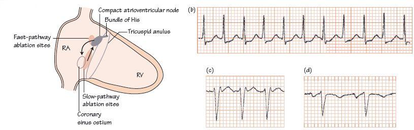
Fig. 45.3 Supraventricular tachycardia (SVT) due to atrioventricular nodal re-entrant tachycardia (AVNRT). Tachycardia, rate about 210 b/min. No P waves before the QRS complex, lead V1 shows R′ (see Fig. 45.2c,d)), a hallmark of AVNRT. The QRS complexes are otherwise normal, but there is widespread ST depression anterolaterally. The heart rate is so high that the ST depression does not necessarily indicate coronary disease. The arrhythmia terminated with adenosine.

Atrioventricular nodal re-entrant tachycardia (AVNRT) is common. Seventy per cent of patients are female; medical help is most commonly sought in those 30–50 years old. In AVNRT an abnormal extra pathway, congenital in origin, near or within the atrioventricular (AV) node allows, from time to time, the depolarizing wave to become trapped within structures local to the AV node, describing a continuous circular re-entrant pattern (Fig. 45.1
Stay updated, free articles. Join our Telegram channel

Full access? Get Clinical Tree


