Fig. 9.1
Paroxysmal atrial fibrillation in a 17-year-old boy. Note the low amplitude fast waveforms (atrial fibrillatory waves) between the “irregularly irregular” QRS intervals. There are a few different QRS morphologies due to aberrant conduction. The atrial fibrillation spontaneously terminates (bottom tracing) as it frequently does in the paroxysmal form in patients without structural heart disease
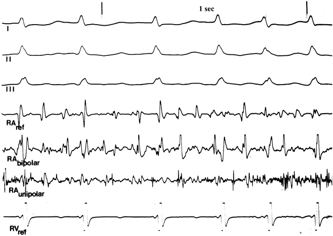
Fig. 9.2
Three electrocardiographic leads and four intracardiac electrograms [three in the right atrium (RA) and one from the right ventricular apex (RV)] illustrating the multi-phasic variable amplitude of the atrial fibrillation wave fronts
Incidence
Atrial fibrillation is the most common rhythm disorder seen in man; prevalence in the general population is estimated at 0.4 %. There is a significant age-related distribution of the arrhythmia with 6 % of the population greater than 80 years of age suffering from atrial fibrillation. Atrial fibrillation remains an extremely rare arrhythmia in the pediatric population. Interestingly, genetic mutations in the KCNQ1 channel have been associated with atrial fibrillation as has the mutation related to the short QT syndrome (Figs. 9.3 and 9.4) has been reported (Chap. 18). Familial atrial fibrillation has involved an addition of nine different genetic defects from 13 loci; many of the genes are implicated in other cardiac arrhythmias (Chap. 19).
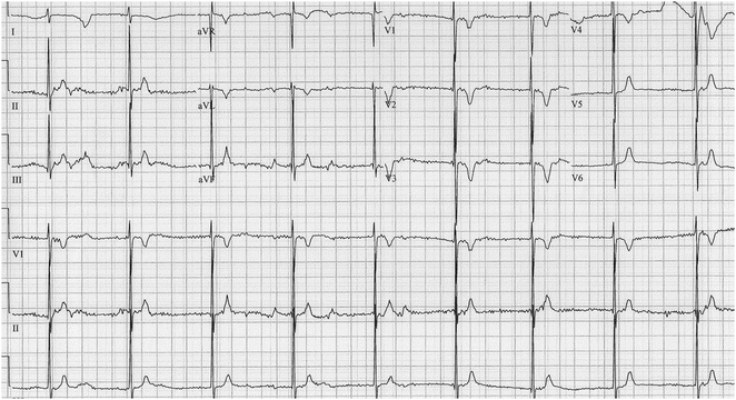
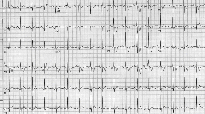
Fig. 9.3
One-week-old girl with congenital atrial fibrillation—the baseline shows a fine fibrillatory waveform, confirmed by esophageal electrogram (not shown)
Fig. 9.4
Same patient as in Fig. 9.3—now in sinus rhythm (only for several hours; usual rhythm is atrial fibrillation). Note the very short QT interval (see text)
Associated Disease
Atrial fibrillation commonly occurs as a comorbid condition with other cardiovascular abnormalities. It is found in patients with congestive heart failure, mitral valve stenosis and insufficiency, hypertension, hyperthyroidism, and some forms of repaired congenital heart malformations. In addition, it is well recognized in the child and young adult with complex, functionally impaired congenital heart malformation, particularly those with extensive atrial incisions or suture lines (Figs. 9.5 and 9.6). Atrial fibrillation is also associated with Wolff–Parkinson–White syndrome. Atrial fibrillation occurring in conjunction with Wolff–Parkinson–White syndrome is a potentially life-threatening situation due to the lack of decremental conduction through the accessory pathway. Some accessory pathways are capable of rapidly conducting the atrial signals to the ventricle leading to ventricular fibrillation (Chaps. 4 and 21). Following elimination of the accessory pathway either surgically or via radiofrequency ablation, atrial fibrillation resolves in a large portion of these patients. Atrial fibrillation also may be associated with several other forms of supraventricular tachycardia in young patients. Our experience and that of others has been that young patients presenting with atrial fibrillation will frequently have other underlying mechanisms of SVT such as AVNRT, AVRT, ectopic atrial tachycardia, or atrial flutter. Ablation of these other tachycardia substrates results in the resolution of atrial fibrillation. Conversion of these forms of SVT with adenosine may result in transient atrial fibrillation (Fig. 9.7) as adenosine shortens atrial refractoriness.
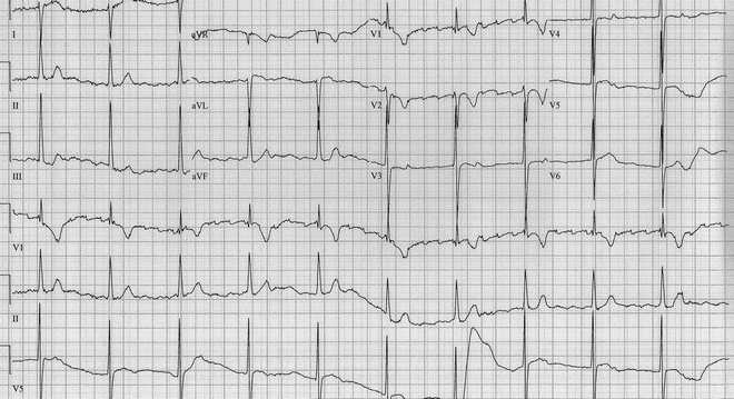
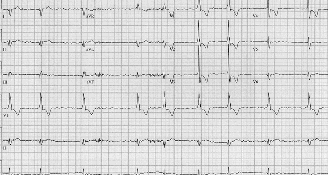
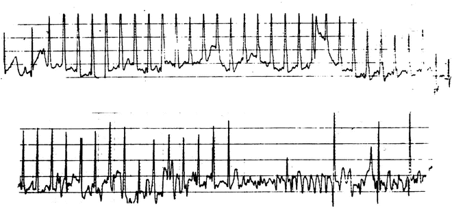
Fig. 9.5
Lead ECG in a 14 months boy after the double switch operation for congenitally corrected transposition of the great arteries. Note the atrial flutter-fibrillatory baseline
Fig. 9.6
12 Lead ECG from a 30-year-old man following the Mustard operation for d-transposition of the great arteries. Note the fine fibrillatory baseline and the “irregular irregularity” of the ventricular response
Fig. 9.7
Six hundred gram premature infant with SVT administered 120 mcg of adenosine. Note: The termination of the SVT but emergence of atrial fibrillation. The fibrillation spontaneously ceased several seconds later
Another interesting association of atrial fibrillation is with obesity. This link has been demonstrated in adults as well as with pediatric patients. The exact physiological mechanism for this connection is unknown currently. We have also seen patients with atrial fibrillation that has probably resulted from enhanced vagal tone. Very strong vagal input can significantly shorten the refractory period of atrial myocardium making the atria more susceptible to microreentrant circuits which underlie atrial fibrillation (see below). This is the physiological mechanism which leads to atrial fibrillation after adenosine administration (Fig. 9.7).
< div class='tao-gold-member'>
Only gold members can continue reading. Log In or Register to continue
Stay updated, free articles. Join our Telegram channel

Full access? Get Clinical Tree


