Atrial Fibrillation
Melvin M. Scheinman, MD
 ESSENTIALS OF DIAGNOSIS
![]() Irregularly irregular ventricular rhythm.
Irregularly irregular ventricular rhythm.
![]() Absence of P waves on the electrocardiogram.
Absence of P waves on the electrocardiogram.
 General Considerations
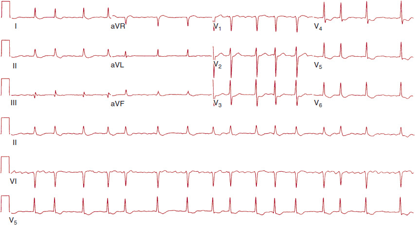
Atrial fibrillation, the most common sustained clinical arrhythmia, is diagnosed by finding an irregularly irregular ventricular rhythm without discrete P waves (Figure 12–1). The QRS complex is usually narrow, but it may be wide if aberrant conduction or bundle branch block is present. Atrial fibrillation associated with the Wolff-Parkinson-White syndrome may occur with very rapid ventricular rates and may be life-threatening. This arrhythmia is diagnosed by its very rapid irregular rate associated with wide preex-cited QRS complexes and requires emergency treatment (see later section, Long-Term Approach).

![]() Figure 12–1. The 12-lead electrocardiogram shows the typical rapid irregular rhythm seen with atrial fibrillation.
Figure 12–1. The 12-lead electrocardiogram shows the typical rapid irregular rhythm seen with atrial fibrillation.
Approximately 4% of the population over age 60 years has sustained an episode of atrial fibrillation, with a particularly steep increase in prevalence after the seventh decade of life. Risk factors for development of atrial fibrillation include heart failure, hypertensive cardiovascular disease, coronary artery disease, and valvular heart disease. Moreover, both sustained and paroxysmal atrial fibrillation have important implications for the development of a cerebrovascular accident (CVA) or other systemic emboli. It is estimated that 15–20% of CVAs in nonrheumatic patients are due to atrial fibrillation.
 Clinical Findings
A. Symptoms & Signs
When called on to manage new-onset atrial fibrillation, it is important to establish the precipitating factors because the type of associated condition determines long-term prognosis. In some patients, episodes of atrial fibrillation may be initiated by caffeine, alcohol, or marijuana use. Atrial fibrillation may result from acute intercurrent ailments. For example, this arrhythmia may develop in patients with hyperthyroidism or lung disease, or after either cardiac or pulmonary surgery, especially in older patients. Atrial fibrillation is also seen in patients with acute pulmonary embolism, myocarditis, or acute myocardial infarction, particularly when the last condition is complicated by either occlusion of the right coronary artery or heart failure. When atrial fibrillation occurs in these settings, it almost always abates spontaneously if the patient recovers from the underlying problem. Hence, management usually involves administration of drugs to control the heart rate, and long-term antiarrhythmic therapy is generally not needed.
Alternatively, atrial fibrillation may occur in association with structural cardiac disease. Important associated conditions include rheumatic mitral stenosis, hypertension, hypertrophic cardiomyopathy, or chronic heart failure. In contrast to patients with acute intercurrent ailments, those with structural heart disease may expect (even with antiar-rhythmic therapy) many recurrences, and chronic atrial fibrillation may supervene.
Lone fibrillation is the term used to describe patients with atrial fibrillation not associated with known cardiac conditions or noncardiac precipitants. The natural history of the atrial fibrillation for those with lone atrial fibrillation is similar to that in patients with structural cardiac disease, in that episodes of atrial fibrillation are likely to recur and, eventually, the arrhythmia may become sustained.
B. Initial Evaluation
The initial evaluation is mainly directed at diagnosing associated conditions (eg, valvular heart disease, hypertension, evidence of heart failure). The cardiac examination during atrial fibrillation will show an irregularly irregular rhythm, variability of the first heart sound, and augmentation of a systolic murmur after long pauses. The initial evaluation of new-onset atrial fibrillation also includes a detailed history focusing on possible precipitating factors as well as the presence of organic cardiac disease. As such, the initial evaluation includes, at a minimum, a careful physical examination, 12-lead electrocardiogram (ECG), chest radiograph, echocardiogram, and tests of thyroid function. Further testing will depend on various aspects of the history or physical examination. Ambulatory ECG monitoring may be helpful in diagnosing the patient with paroxysmal atrial fibrillation. For example, if atrial fibrillation is usually precipitated by exercise, then an exercise treadmill test is appropriate. In the patient with frequent episodes of paroxysmal atrial fibrillation, a 24- to 48-hour ambulatory ECG recording may discern whether atrial fibrillation was triggered by another arrhythmia, such as a premature atrial complex alone, or whether the fibrillation was preceded by an episode of supraventricular tachycardia. In addition, patients with vagally mediated fibrillation will typically have episodes either after heavy meals or during sleep. These clues may help identify those patients who may respond to specific approaches (see later section, Treatment).
 Differential Diagnosis
The ECG diagnosis of atrial fibrillation is usually straightforward and is characterized by an irregularly irregular rhythm with a narrow QRS complex unless there is associated bundle branch block. The chief diagnostic problem relates to patients showing short bursts of irregular atrial tachycardia (which are common on random ambulatory ECG recordings in the elderly). In addition, patients with atrial flutter may have varying atrioventricular (AV) conduction, which may serve to mimic atrial fibrillation. Careful evaluation of all leads looking for the characteristic flutter pattern is important. Less commonly, patients presenting with multifocal atrial tachycardia may mimic the irregularity found in those with atrial fibrillation. Careful analysis in the latter group shows an irregular atrial tachycardia with at least three P-wave morphologies. A very uncommon arrhythmia that may mimic atrial fibrillation is seen in patients with dual AV nodal physiology with dual conduction over the fast and slow AV nodal pathways. For this diagnosis, identify that a single sinus P wave is associated with two QRS complexes. These patients are readily cured with slow pathway ablation.
 Treatment
The objectives of therapy include (1) achieving rate control, (2) restoring sinus rhythm (where feasible), and (3) decreasing the risk of CVA. The principles of treatment discussed in this chapter largely follow those promulgated in the recent American College of Cardiology (ACC)/American Heart Association (AHA)/European Society of Cardiology (ESC) guidelines.
European Heart Rhythm Association, et al. ACC/AHA/ESC 2006 guidelines for the management of patients with atrial fibrillation—executive summary: a report of the American College of Cardiology/American Heart Association Task Force on Practice Guidelines and the European Society of Cardiology Committee for Practice Guidelines (Writing Committee to Revise the 2001 Guidelines for the Management of Patients with Atrial Fibrillation). J Am Coll Cardiol. 2006;48(4):854–906. [PMID: 16904574]
Wann SL, et al. 2011 ACCF/AHA/HRS focused update on the management of patients with atrial fibrillation (updating the 2006 guideline); a report of the American College of Cardiology/American Heart Association Task Force on Practice Guidelines. J Am Coll Cardiol. 2011;57(2):223–42. [PMID: 21177058]
A. Rate Control
If the patient has atrial fibrillation and a rapid rate associated with severe heart failure or cardiogenic shock, emergency direct-current cardioversion is indicated. For patients with atrial fibrillation associated with rapid rate but with stable hemodynamics, attempts to achieve acute rate control are indicated. Drugs to slow the ventricular rate in patients with atrial fibrillation (Table 12–1) include digitalis preparations, calcium channel blockers (verapamil or diltiazem), and β-blockers. If rapid rate control is desired, then calcium channel blockers and β-blockers are far more effective than digitalis. In addition, a common misconception is that digitalis therapy is associated with acute conversion to sinus rhythm, but carefully controlled studies have shown that conversion to sinus rhythm is no more likely with digoxin than with placebo. As emphasized later, digitalis and intravenous calcium channel blocker therapy are contraindicated in patients with Wolff-Parkinson-White syndrome and atrial fibrillation. Intravenous diltiazem has been shown to be safe and effective for patients with atrial fibrillation and a modest degree of heart failure. In patients refractory to these agents, intravenous amiodarone may be effective but requires many hours before rate control is achieved. In patients with a known history of congestive heart failure, use of intravenous β-blockers or calcium channel blockers may aggravate cardiac failure. In this subset, digitalis or intravenous amiodarone would be the preferred agent for rate control.
Table 12–1. Intravenous Pharmacologic Agents for Heart Rate Control in Atrial Fibrillation

B. Long-Term Antiarrhythmic Therapy and Elective Cardioversion
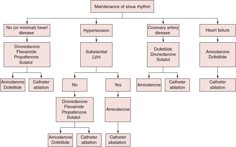
For patients who have had a single, initial episode of atrial fibrillation with no significant hemodynamic problems, no specific therapy is required because repeat episodes may not occur for many years. In contrast, patients who manifest frequent recurrences may be candidates for long-term anti-arrhythmic therapy with class Ia (quinidine, procainamide, and disopyramide), class Ic (propafenone and flecainide), or class III (sotalol, amiodarone, dronedarone, and dofetilide) agents, all of which are more effective than placebo in maintaining sinus rhythm (Figure 12–2).

![]() Figure 12–2. Therapy to maintain sinus rhythm in patients with recurrent paroxysmal or persistent atrial fibrillation. Drugs are listed alphabetically and not in order of suggested use. The seriousness of heart disease progresses from left to right, and selection of therapy in patients with multiple conditions depends on the most serious condition present. LVH, left ventricular hypertrophy. (Reproduced, with permission, from Wann SL, et al. J Am Coll Cardiol. 2011;57[2]:223–42.)
Figure 12–2. Therapy to maintain sinus rhythm in patients with recurrent paroxysmal or persistent atrial fibrillation. Drugs are listed alphabetically and not in order of suggested use. The seriousness of heart disease progresses from left to right, and selection of therapy in patients with multiple conditions depends on the most serious condition present. LVH, left ventricular hypertrophy. (Reproduced, with permission, from Wann SL, et al. J Am Coll Cardiol. 2011;57[2]:223–42.)
Stay updated, free articles. Join our Telegram channel

Full access? Get Clinical Tree


