Fig. 10.1
12-Lead ECG from a 9-month-old boy with atrial ectopic tachycardia at 150 bpm. There is variable A-V conduction. The P-wave is large in many leads and has a leftward inferior (positive in leads I, II, aVF) but anterior (positive in lead V1) axis suggesting a high left atrial origin near the right upper pulmonary vein
Noninvasive testing may clarify the diagnosis (Table 10.1). In contrast to reentrant forms of SVT, Holter monitoring may demonstrate a characteristic gradual acceleration of the heart rate in AET, the so-called warm-up period. The first beat of the tachycardia typically occurs late in the cycle, and the initial P-wave will have the same morphology as subsequent P-waves. Participation of the ventricle in the tachycardia is not required, excluding an accessory pathway in these patients. During periods of reduced adrenergic tone and heightened parasympathetic tone (sleep, carotid sinus massage), the P-waves of an AET may not conduct to the ventricles, resulting in varying degrees of atrioventricular block. On occasion, this block can be induced with adenosine (Fig. 10.2); on the other hand, some AETs may be transiently suppressed with adenosine, limiting the usefulness of this test.
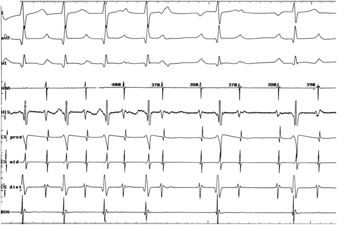
Table 10.1
Criteria for automatic atrial tachycardia
P-wave preceding each QRS |
|---|
P-wave of different morphology from sinus |
Persistence of tachycardia in the presence of AV block |
“Warm-up” period |
Catecholamine sensitive |
Fig. 10.2
Atrioventricular block with adenosine during AAT. The atrial tachycardia persists at a cycle length of 370–390 ms despite AV block induced by adenosine. In addition to demonstrating that the ventricular is not a participant in the tachycardia, the P-wave, which had been buried within the preceding T-wave, becomes clearly discernable
AET also has distinguishing features observed at electrophysiology study. First, it cannot be terminated by overdrive pacing. If ventricular participation can be excluded during either spontaneous or adenosine-induced AV node Wenckebach, the remaining diagnostic possibility for a narrow QRS tachycardia with P-waves preceding each QRS complex is atrial flutter (intra-atrial reentrant tachycardia). While macroreentrant tachycardia, including atrial flutter, can be interrupted with appropriate atrial overdrive pacing, an automatic tachycardia is only momentarily suppressed before the ectopic focus gradually warms up and returns. Another diagnostic finding is the sequence of atrial-atrial-ventricular electrograms (A-A-V) following cessation of ventricular burst pacing, elicited in almost 80 % of patients with a paroxysmal form of this arrhythmia. Use of catecholamine to induce AET may make it difficult to identify the mechanism of the tachycardia (Fig. 10.3).
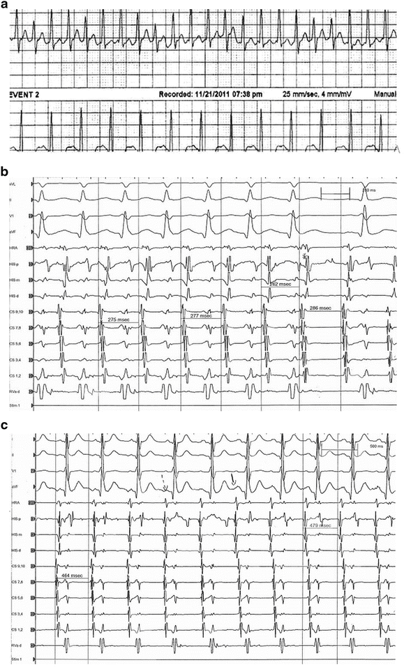
Fig. 10.3
Panel a: Rhythm strip from a 16-year-old with exertional palpitations. Rhythm strip shows narrow QRS tachycardia, with slight cycle length variability, 260–280 ms, and also a suggestion of P-waves. Panel b: Atrial burst pacing on isoproterenol induces clinical tachycardia, initially with 1:1 AV conduction, where P-wave is buried in preceding T-wave. Block of an atrial premature beat #7 (asterisk) suppresses the following atrial beat with identical atrial activation sequences during the tachycardia consistent with an abnormal automatic mechanism and thus eliminates tachycardia mediated by concealed accessory pathway. Panel c: After discontinuing isoproterenol, AET slowing leads to resumption of sinus rhythm, as evidenced by the change in P-wave morphology and atrial activation sequence
Treatment
Treatment presents two options: antiarrhythmic medications or radiofrequency ablation. There are no controlled, randomized series to evaluate drug therapy; available data are difficult to interpret because of selection bias in retrospective studies. Digoxin, propranolol, and verapamil are ineffective. Class III drugs like amiodarone and sotalol, and IC drugs (propafenone and flecainide) have been more successful in small retrospective series. The addition of a beta-blocker to flecainide may be helpful, and in refractory cases, flecainide in combination with amiodarone may be considered. AET may be a complication of transcatheter atrial septal defect closure devices; treatment requires removal of the device; however, the tachycardia may persist and ablation of the identified site be necessary (Fig. 10.4).

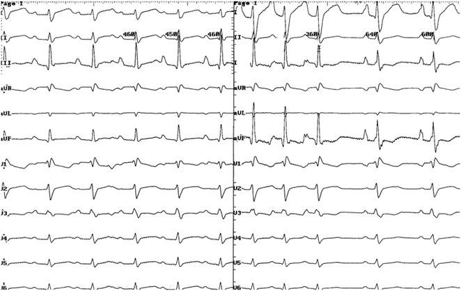
Fig. 10.4
13 Lead ECGs from 5-year-old child, s/p Amplatzer septal occluder for secundum ASD. Panel a: Atrial ectopy following device implant subsequently progressed to non-sustained AET (panel b) and the development of mild LV systolic dysfunction. Pharmacologic treatment was ineffective, which led 1 year later to surgical explant of the device and patch closure of the ASD. Panel c: AET became almost incessant, prompting catheter radiofrequency ablation. Lesions on the left side at the lower margin of the patch slowed the tachycardia, and subsequent lesions on the right side of the patch, near the CS os terminated the tachycardia. LV function subsequently normalized
The acute success rate of radiofrequency ablation for AET approaches 95–100 %. Experience in children demonstrates that a majority of the foci are in the left atrium, particularly near the right pulmonary veins and in the atrial appendage. In contrast, the anatomic distribution in adult patients is predominantly on the right side, particularly near the SVC/RA junction, right atrial appendage, and the coronary sinus os. When the focus (i) in children is on the right side, they cluster in and around the right atrial appendage. The technical aspects of successful ablation include identification of the earliest intra-atrial electrogram preceding the onset of the surface P-wave by at least 20 ms, pace-mapping to assure that P-waves originating from the pacing site are identical to those of the AAT and computer-based intra-cardiac mapping techniques (Figs. 10.5 and 10.6).
 < div class='tao-gold-member'>


Only gold members can continue reading. Log In or Register to continue
Stay updated, free articles. Join our Telegram channel
Full access? Get Clinical Tree
