Fig. 4.1
The left panel is a diagrammatic representation of pulmonary cavities on each side of the thorax with the mediastinum in between. The right panel illustrates the divisions of the mediastinum. Figure adapted from Grant’s Dissector, 12th edn. by EK Sauerland (Fig. 1.14, left; Fig. 1.24, right)
The inferior aperture of the thorax is formed by the lower margin of the ribs and costal cartilages and is closed off from the abdomen by the respiratory diaphragm (Fig. 4.1). The superior aperture of the thorax leads to the neck and the upper extremity; it is formed by the first ribs and their articulation with the manubrium and first thoracic vertebra. The superior aperture of the thorax, or root of the neck, allows for the passage of structures between the neck and thoracic cavity. The clavicle crosses the first rib at its anterior edge close to its articulation with the manubrium. Structures exiting the superior thoracic aperture and communicating with the upper extremity pass between the first rib and clavicle. Anatomists often refer to the superior thoracic aperture as the thoracic inlet due to the entrance of air and food through the trachea and esophagus, respectively. However, clinicians may refer to the superior thoracic aperture as the thoracic outlet due to the fact that arteries and nerves leave the thorax through this area to enter the neck and upper extremities.
4.3 Bones of the Thoracic Wall
4.3.1 The Thoracic Cage
The skeleton of the thoracic wall is composed of the 12 pairs of ribs, the thoracic vertebra (and intervertebral disks), and the sternum. Articulating with the thorax are the bones of the pectoral girdle, the clavicle and the scapula (Fig. 4.2). Nerves and blood vessels entering the upper extremity pass between the clavicle and first rib.
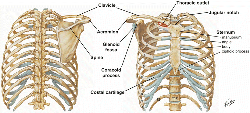

Fig. 4.2
The left panel illustrates the bones of the thorax from an anterior view. The right panel is a posterior view of the bony thorax. ©1998 Elsevier Inc. All rights reserved. www.netterimages.com, Frank Netter
The 12 thoracic vertebrae, which comprise the midline of the posterior wall of the thorax, articulate with the 12 pairs of ribs. Each thoracic vertebra has a body anteriorly and a neural arch which form a vertebral foramen. Extending from the neural arch posteriorly is an elongated, inferiorly slanting, spinous process and extending bilaterally are transverse processes. The transverse processes of thoracic vertebrae become progressively shorter from superior to inferior. The parts of the neural arch between the body and transverse processes are the pedicles , while the parts between the transverse processes and spinous process are the laminae (Fig. 4.3). Thoracic vertebrae have several articular surfaces, called facets . Superior and inferior articular facets form facet joints with vertebrae above and below, respectively. Costal facets are sites of articulation with ribs. Generally, each rib articulates with two adjacent vertebrae such that the head fits between adjacent bodies and the tubercle (slightly distal to the neck of the rib) articulates with the transverse process of the vertebra below. Therefore, a rib articulates with its corresponding thoracic vertebrae through the body and transverse process and with the vertebra above through its body. The articular surfaces on transverse processes are called costal facets , while those on the bodies, which generally share rib articulation with the vertebra above, are referred to as costal demifacets .
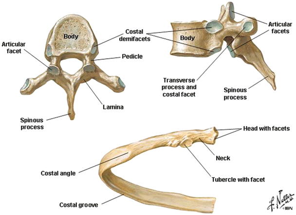

Fig. 4.3
The T6 vertebra as viewed from above (upper left) and laterally (upper right), and a typical rib (lower). ©1998 Elsevier Inc. All rights reserved. www.netterimages.com, Frank Netter
There are a few exceptions to this general relationship between thoracic vertebrae and ribs. The first thoracic vertebra shares the articulation with the second rib, but receives all of the articulation of the first rib superiorly. Ribs 10, 11, and 12 only articulate with their corresponding vertebrae.
The ribs form the largest part of the bony wall of the thorax (Fig. 4.2). Each rib articulates with one or two thoracic vertebrae, and the upper ten ribs articulate directly or indirectly with the sternum anteriorly. The upper seven ribs are referred to as true ribs because each connects to the sternum via its own costal cartilage. Ribs 8–10 are referred to as false ribs because they connect to the sternum (indirectly) by joining the articular cartilage of the seventh rib. Ribs 11 and 12 are referred to as floating ribs because they do not connect to the sternum, but end in the musculature of the abdominal wall. Each rib has a head that articulates with the thoracic vertebra and a relatively thin flat shaft that is curved (Fig. 4.3). The costal angle, the sharpest part of the curved shaft, is located where the rib turns anteriorly. At the inferior margin of the shaft, the internal surface of each rib is recessed to form a costal groove. This depression provides some protection to the intercostal neurovascular bundle, something that must be considered when designing devices for intercostal access to the thorax. The heads of ribs 2–9 have two articular facets for articulation with the vertebra of the same level and the vertebra above. The heads of ribs 1, 10, 11, and 12 only articulate with the vertebra of the same number and, consequently, have only one articular facet. In ribs 1–10, the head is connected to the shaft by a narrowing called the neck. At the junction of the head and the neck is a tubercle that has an articular surface for articulation with the costal facet of the transverse process. Ribs 11 and 12 do not articulate with the transverse process of their respective vertebra and do not have a tubercle or, therefore, a defined neck portion.
The sternum is the flat bone making up the median anterior part of the thoracic cage (Fig. 4.2). It is composed of three parts—the manubrium, body, and xiphoid process. The manubrium (from the Latin word for handle, like the handle of a sword) is the superior part of the sternum; it is the widest and thickest part of the sternum. The manubrium alone articulates with the clavicle and the first rib. The sternal heads of the clavicle can be readily seen and palpated at their junction with the manubrium. The depression between the sternal heads of the clavicle above the manubrium is the suprasternal, or jugular, notch. The manubrium and the body of the sternum lie in slightly different planes and thus form a noticeable and easily palpated angle, the sternal angle (of Louis), at the point where they meet. The second rib articulates with the body of the sternum and the manubrium at the sternal angle. The body of the sternum is formed from the fusion of segmental bones (the sternebrae). The remnants of this fusion may be seen in the transverse ridges of the sternal body, especially in young people. The third through sixth ribs articulate with the body of the sternum, and the seventh rib articulates at the junction of the body and xiphoid process. The xiphoid process is the inferior-most part of the sternum and is easily palpated. It lies at the level of T9–T10 vertebrae and marks the inferior boundary of the thoracic cavity anteriorly. It also lies at the level even with the central tendon of the diaphragm and the inferior border of the heart. When a median sternotomy is performed on a patient, typically a sternal saw is used to cut through the manubrium, the sternal body, and the xiphoid process. For more details, refer to Chap. 33.
4.3.2 The Pectoral Girdle
Many of the muscles encountered on the wall of the anterior thorax are attached to the bones of the pectoral girdle and the upper extremity. Since movement of these bones can impact the anatomy of vascular structures communicating between the thorax and upper extremity, it is important to include these structures in a discussion of the thorax.
The clavicle is a somewhat “S”-shaped bone that articulates at its medial end with the manubrium of the sternum and at its lateral end with the acromion of the scapula (Fig. 4.2). It is convex medially and concave laterally. The scapula is a flat triangular bone, slightly concave anteriorly, that rests upon the posterior thoracic wall. It has a raised ridge posteriorly called the spine that ends in a projection of bone called the acromion that articulates with the clavicle. The coracoid process is an anterior projection of bone from the superior border of the clavicle that serves as an attachment point for muscles that act on the scapula and upper extremity. The head of the humerus articulates with the shallow glenoid fossa of the scapula forming the glenohumeral joint. The clavicle serves as a strut to hold the scapula in position away from the lateral aspect of the thorax. It is a highly mobile bone, with a high degree of freedom at the sternoclavicular joint that facilitates movement of the shoulder girdle against the thorax. The anterior extrinsic muscles of the shoulder pass from the wall of the thorax to the bones of the shoulder girdle.
4.4 Muscles of the Thoracic Wall
4.4.1 The Pectoral Muscles
Several muscles of the thoracic wall, including the most superficial ones that create some of the contours of the thoracic wall, are muscles that act upon the upper extremity. Some of these muscles form important surface landmarks on the thorax, and others have relationships to vessels that communicate with the thorax. In addition to moving the upper extremity, some of these muscles also can play a role in movement of the thoracic wall and participate in respiration. The pectoralis major muscle forms the surface contour of the upper lateral part of the thoracic wall (Fig. 4.4). It originates on the clavicle (clavicular head), the sternum, and ribs (sternocostal head), and inserts near the greater tubercle of the humerus. The lower margin of this muscle, passing from the thorax to the humerus, forms the major part of the anterior axillary fold. The pectoralis major muscle is a powerful adductor and medial rotator of the arm.
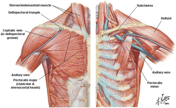

Fig. 4.4
The musculature of the anterior thoracic wall . The left panel shows the superficial muscles intact. The left panel shows structures deep to the pectoralis major muscle. ©1998 Elsevier Inc. All rights reserved. www.netterimages.com, Frank Netter
The pectoralis minor muscle is a much smaller muscle and lies immediately deep to the pectoralis major muscle (Fig. 4.4). It originates on ribs 3–5 and inserts upon the coracoid process of the scapula. This muscle forms part of the anterior axillary fold medially. It acts to depress and stabilize the scapula when upward force is exerted on the shoulder.
The anterior part of the deltoid muscle also forms a small aspect of the anterior thoracic wall. This muscle has its origin on the lateral part of the clavicle and the acromion and spine of the scapula (Fig. 4.4). It inserts upon the deltoid tubercle of the humerus and is the most powerful abductor of the arm. The anterior deltoid muscle borders the superior aspect of the pectoralis major muscle. The depression found at the junction of these two muscles is called the deltopectoral groove . Importantly, within this groove, the cephalic vein can consistently be found. The muscles diverge at their origins on the clavicle, creating an opening bordered by these two muscles and the clavicle known as the deltopectoral triangle . Through this space the cephalic vein passes to join the axillary vein.
The subclavius is a small muscle originating on the lateral inferior aspect of the clavicle and inserting on the sternal end of the first rib (Fig. 4.4). This muscle depresses the clavicle and exerts a medial traction on the clavicle that stabilizes the sternoclavicular joint. In addition to these actions, the subclavius muscle provides a soft surface on the inferior aspect of the clavicle that serves to cushion the contact of this bone with structures passing under the clavicle (i.e., nerves of the brachial plexus and the subclavian artery) when the clavicle is depressed during movement of the shoulder girdle and especially when the clavicle is fractured.
The serratus anterior muscle originates on the lateral aspect of the first eight ribs and inserts upon the medial aspect of the scapula (Fig. 4.5). This muscle forms the serrated contour of the lateral thoracic wall in individuals with good muscle definition. The serratus anterior forms the medial border of the axilla and acts to pull the scapula forward (protraction) and to stabilize the scapula against a posterior force on the shoulder; it also assists with cranial rotation of the scapula with elevation.
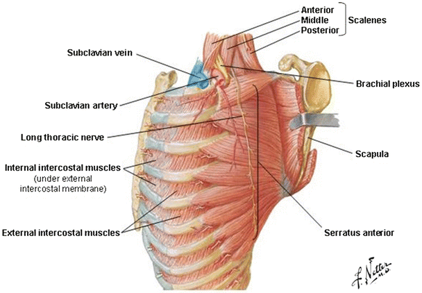

Fig. 4.5
A lateral view of the musculature of the thoracic wall. ©1998 Elsevier Inc. All rights reserved. www.netterimages.com, Frank Netter
4.4.2 The Intercostal Muscles
Each rib is connected to the one above and below by a series of three intercostal muscles. The external intercostal muscles are the most superficial (Fig. 4.5). These muscles course in an obliquely medial direction as they pass from superior to inferior between the ribs. Near the anterior end of the ribs, the external intercostal muscle fibers are replaced by the external intercostal membrane. Deep to the external intercostals are the internal intercostals (Figs. 4.5 and 4.6). The direction of the internal intercostal muscle fibers is perpendicular to the external intercostals. Near the posterior end of the ribs, the internal intercostal muscle fibers are replaced by the internal intercostal membrane. The deepest layer of intercostal muscle is the innermost intercostal muscle (Fig. 4.6). These muscles have a fiber direction similar to the internal intercostals, but they form a separate plane. The innermost intercostal muscles become membranous near the anterior and posterior ends of the ribs. The intercostal nerves and vessels pass between the internal and innermost intercostal muscles, which help to distinguish between the two muscular layers. There are two additional sets of muscles in the same plane as the innermost intercostals—the subcostals and the transversus thoracic muscles. The subcostal muscles are located posteriorly and span more than one rib. The transversus thoracic muscles are found anteriorly and pass from the internal surface of the sternum to ribs 2–6, extending superiorly and laterally (Fig. 4.6).
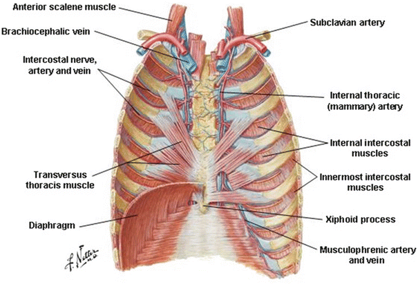

Fig. 4.6
The deep musculature of the anterior thoracic wall viewed from the posterior side. ©1998 Elsevier Inc. All rights reserved. www.netterimages.com, Frank Netter
The intercostal muscles, especially the external and internal intercostals, are involved with respiration by elevating or depressing the ribs. The external intercostal muscles and the anterior interchondral part of the internal intercostals act to elevate the ribs. The interosseous parts of the internal intercostal muscles depress the ribs. The innermost intercostals likely have an action similar to the internal intercostals. The subcostal muscles probably help to elevate the ribs. The action of transversus thoracis is not well understood. It may, by virtue of its attachments, help to depress the ribs with forced exhalation or play a role in proprioception. In so-called less or minimally invasive surgical procedures performed to gain access to the heart, one needs to transect through the intercostal muscles (see Chap. 35).
4.4.3 Respiratory Diaphragm
The respiratory diaphragm is the musculotendinous sheet separating the abdominal and thoracic cavities (Figs. 4.6 and 4.7). It is considered the primary muscle of respiration. The diaphragm originates along the inferior border of the rib cage, the xiphoid process of the sternum, the posterior abdominal wall, and the upper lumbar vertebra. The medial and lateral arcuate ligaments are thickenings of the investing fascia over the quadratus lumborum (lateral) and the psoas major (medial) muscles of the posterior abdominal wall that serve as attachments for the diaphragm (Fig. 4.7). The right and left crura of the diaphragm attach to lumbar vertebrae. They originate on the bodies of lumbar vertebrae 1–3, their intervertebral disks, and the anterior longitudinal ligament spanning these vertebrae. The diaphragm ascends from its origin to form a right and left dome, the right dome being typically higher than the left. The muscular part of the diaphragm is dome-shaped at rest. Contraction (during inhalation) causes the dome of the diaphragm to flatten (downward), increasing the volume of the thoracic cavity. The aponeurotic central part of the diaphragm, called the central tendon , contains the opening for the inferior vena cava (Fig. 4.7). The esophagus also passes through the diaphragm, and the hiatus for the esophagus is created by a muscular slip originating from the right crus of the diaphragm. The aorta passes from the thorax to the abdomen behind the diaphragm, under the median arcuate ligament created by the intermingling of fibers from the right and left crura of the diaphragm. The inferior vena cava, esophagus, and aorta pass from the thorax to the abdomen at thoracic vertebral levels 8, 10, and 12, respectively.
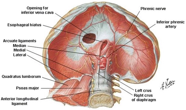

Fig. 4.7
The abdominal side of the respiratory diaphragm illustrating the origins of the muscle. ©1998 Elsevier Inc. All rights reserved. www.netterimages.com, Frank Netter
4.4.4 Other Muscles of Respiration
The scalene muscles and the sternocleidomastoid muscle in the neck also contribute to respiration, especially during deep inhalation (Figs. 4.4 and 4.5). Collectively, the scalene muscles have their origin on the transverse processes of cervical vertebra 2–7. The anterior and middle scalenes insert on the first rib and the posterior scalene on the second rib. As its name suggests, the sternocleidomastoid originates on the sternum and medial clavicle and inserts on the mastoid process of the skull. Although the primary action of sternocleidomastoid is on the head and neck, it is also capable of exerting an upward force on the thorax with forced respiration. The muscles of the anterior abdominal wall are also involved with respiration. These muscles, the rectus abdominis, external and internal abdominal obliques, and the transverses abdominis, act together during forced exhalation to depress the rib cage and to increase intra-abdominal pressure forcing the diaphragm to expand upward, reducing the volume of the pulmonary cavities. The mechanics of respiration are explained in detail in Sect. 4.11.2.
4.5 Nerves of the Thoracic Wall
The wall of the thorax receives its innervation from intercostal nerves (Fig. 4.8). These nerves are the ventral rami of segmental nerves, leaving the spinal cord at the thoracic vertebral levels. Intercostal nerves are mixed nerves, carrying both somatic motor and sensory fibers, as well as autonomics to the skin. The intercostal nerves pass out of the intervertebral foramina and run inferior to the ribs. As they reach the costal angle, the nerves pass between the innermost and the internal intercostal muscles. The motor innervation to all the intercostal muscles comes from the intercostal nerves. These nerves give off lateral and anterior cutaneous branches that provide cutaneous sensory innervation to the skin of the thorax (Fig. 4.8). The intercostal nerves also carry sympathetic nerve fibers to the sweat glands, smooth muscle, and blood vessels. However, the first two intercostal nerves are considered atypical. The first intercostal nerve divides shortly after it emerges from the intervertebral foramen. The larger superior part of this nerve joins the brachial plexus to provide innervation to the upper extremity. The lateral cutaneous branch of the second intercostal nerve is large and typically pierces the serratus anterior muscle to provide sensory innervation to the floor of the axilla and medial aspect of the arm. The nerve associated with the 12th rib is the subcostal nerve and, since there is no rib below this level, is considered a nerve of the abdominal wall.
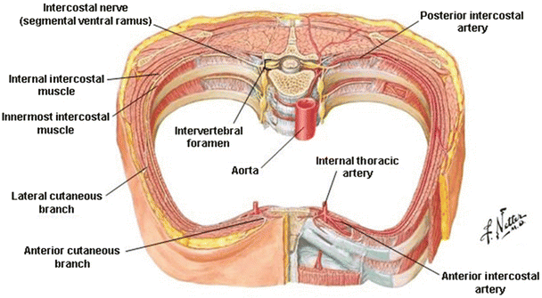

Fig. 4.8
A typical set of intercostals arteries and nerves . ©1998 Elsevier Inc. All rights reserved. www.netterimages.com, Frank Netter
The pectoral muscles receive motor innervation from branches of the brachial plexus of nerves (derived from cervical levels 5–8 and thoracic level 1) which supply the muscles of the shoulder and upper extremity. The lateral and medial pectoral nerves, branches of the lateral and medial cords of the brachial plexus, supply the pectoralis major and minor muscles (Fig. 4.9). The pectoralis major muscle is innervated by both nerves and the pectoralis minor muscle by only the medial pectoral nerve, which pierces this muscle before entering the pectoralis major. The serratus anterior muscle is innervated by the long thoracic nerve which originates from ventral rami of C5, C6, and C7 (Figs. 4.5 and 4.9). The deltoid muscle is innervated by the axillary nerve, a branch of the posterior cord of the brachial plexus (Fig. 4.9). Finally, the subclavius muscle is innervated by its own nerve from the superior trunk of the brachial plexus.
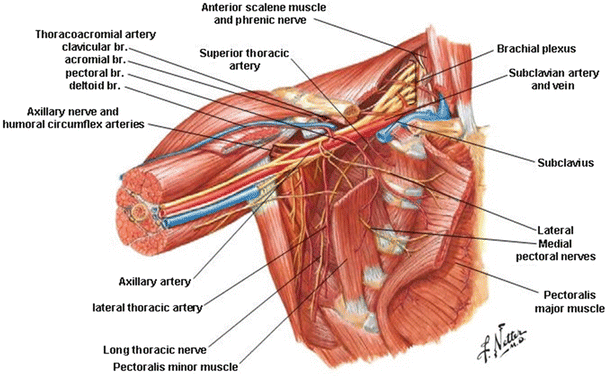

Fig. 4.9
The nerves and arteries of the axilla viewed with the pectoralis major and minor muscles reflected. ©1998 Elsevier Inc. All rights reserved. www.netterimages.com, Frank Netter
4.6 Vessels of the Thoracic Wall
The intercostal muscles and the skin of the thorax receive their blood supply from both the intercostal arteries and the internal thoracic artery (Figs. 4.6 and 4.8). Intercostal arteries 3–11 (and the subcostal artery) are branches directly from the thoracic descending aorta. The first two intercostal arteries are branches of the supreme intercostal artery, which is a branch of the costocervical trunk from the subclavian artery. The posterior intercostals run with the intercostal nerve and pass with the nerve between the innermost and internal intercostal muscles. The intercostals then anastomose with anterior intercostal branches arising from the internal thoracic artery descending lateral to the sternum. The internal thoracic arteries are anterior branches from the subclavian arteries. The anterior and posterior intercostals anastomoses create an anastomotic network around the thoracic wall. The intercostal arteries are accompanied by intercostal veins (Fig. 4.6). These veins drain to the azygos system of veins in the posterior mediastinum. The anatomy of the azygos venous system is described in detail in Sect. 4.10.2. Anteriorly, the intercostal veins drain to the internal thoracic veins which, in turn, drain to the subclavian veins in the superior mediastinum (Fig. 4.7).
The intercostal nerves, arteries, and veins run together in each intercostal space, just inferior to each rib. They are characteristically found in this order (vein, artery, and nerve) with the vein closest to the rib.
The diaphragm receives blood from the musculophrenic arteries and terminal branches of the internal thoracic arteries, which runs along the anterior superior surface of the diaphragm (Fig. 4.6). There is also a substantial blood supply to the inferior aspect of the diaphragm from the inferior phrenic arteries, the superior-most branches from the abdominal aorta that branch along the inferior surface of the diaphragm (Fig. 4.7).
The muscles of the pectoral region get their blood supply from branches of the axillary artery. This artery is the continuation of the subclavian artery emerging from the thorax and passing under the clavicle (Fig. 4.9). The first branch of the axillary artery, the superior (supreme) thoracic artery, supplies blood to the first two intercostal spaces. The second branch forms the thoracoacromial artery or trunk. Subsequently, this artery gives rise to four branches (pectoral, deltoid, clavicular, acromial) that supply blood to the pectoral muscles, the deltoid muscle, the clavicle, and the subclavius muscle. The lateral thoracic artery, the third branch from the subclavian artery, participates along with the intercostal arteries in supplying the serratus anterior muscle. Additional distal branches from the axillary artery, the humeral circumflex arteries, also participate in blood supply to the deltoid muscle. Venous blood returns through veins of the same names to the axillary vein.
4.7 The Superior Mediastinum
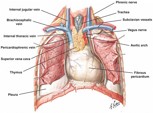
The superior mediastinum is the space behind the manubrium of the sternum (Fig. 4.1). It is bounded by parietal (mediastinal) pleura on each side and the first four thoracic vertebrae behind. It is continuous with the root of the neck at the top of the first ribs and with the inferior mediastinum below the transverse thoracic plane, a horizontal plane that passes from the sternal angle through the space between the T4 and T5 vertebrae. The superior mediastinum contains several important structures including the branches of the aortic arch, the veins that coalesce to form the superior vena cava, the trachea, the esophagus, the vagus and phrenic nerves, the cardiac plexus of autonomic nerves, the thoracic duct, and the thymus (Fig. 4.10).
 < div class='tao-gold-member'>


Only gold members can continue reading. Log In or Register to continue
Stay updated, free articles. Join our Telegram channel

Full access? Get Clinical Tree


