Fig. 5.1
Position of the heart in the thorax. The heart lies in the protective thorax, posterior to the sternum and costal cartilages, and rests on the superior surface of the diaphragm. The heart assumes an oblique position in the thorax, with two-thirds to the left of midline. It is located between the two lungs which occupy the lateral spaces called the pleural cavities. The space between these two cavities is referred to as the mediastinum. The heart lies obliquely in a division of this space, the middle mediastinum, surrounded by the pericardium. Marieb, Elaine N.: Wilhelm, Paticia Brady; Mallatt, Jon B., Human Anatomy, 7th, © 2013. Printed and electronically reproduced by permission of Pearson Education, Inc., New York, New York
The mediastinum is divided first into the superior and inferior mediastinum by a midsagittal imaginary line called the transverse thoracic plane . This plane passes through the sternal angle (junction of the manubrium and body of the sternum) and the space between thoracic vertebrae T4 and T5. This plane acts as a convenient landmark as it also passes through the following structures: the bifurcation of the trachea, the superior border of the pericardium, the artificial division of the ascending and arch of the aortic artery, and the bifurcation of the pulmonary trunk into the pulmonary arteries.
The human heart assumes an oblique position in the thorax, with two-thirds to the left of midline (Figs. 5.2 and 5.3). The heart is roughly in a plane that runs from the right shoulder to the left nipple. The base is located below the 3rd rib as it approaches the sternum (note that the sternal angle occurs at the level of the 2nd rib). The base is directed superiorly, to the right of midline, and posterior. The pointed apex projects to the left of midline and anterior. Thus, the heartbeat can be easily palpated between the 5th and 6th ribs (just inferior to the left nipple) from the apex of the heart where it comes into close proximity of the thoracic wall. Importantly, the heart lies in such an oblique plane that it is often referred to as being horizontal. Thus, the anterior side may be imagined as the superior and the posterior side as inferior (for additional detail on attitudinally correct cardiac anatomy, see Chap. 2).
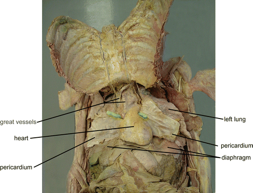
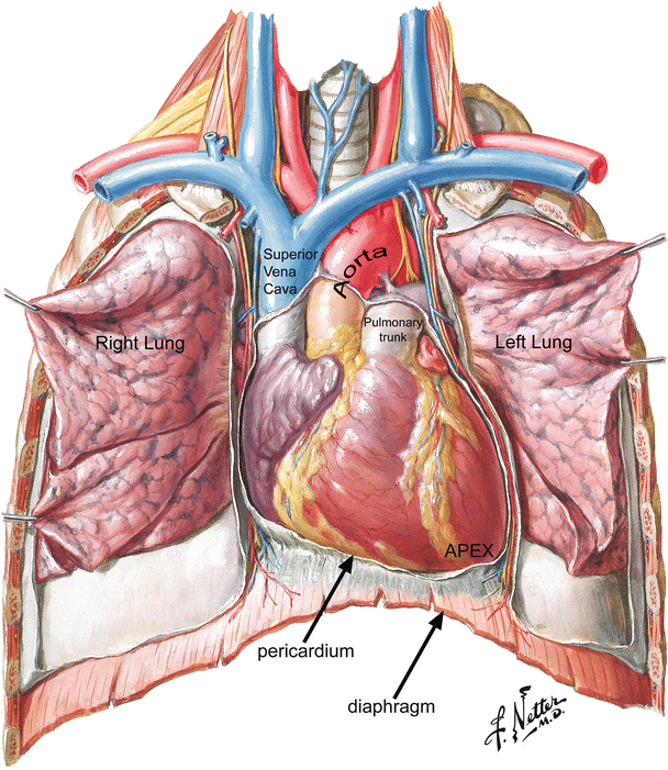
Fig. 5.2
Human cadaver dissection in which the ribs were cut laterally and the sternum and ribs reflected superiorly. This dissection exposes the contents of the thorax (the heart, great vessels, lungs, and diaphragm)
Fig. 5.3
The anterior surface of the heart. The atria are positioned superior to (posterior to) and to the right of their respective ventricles. From superior to inferior, down the anterior surface of the heart, runs the anterior interventricular sulcus (“a groove”). This sulcus separates the left and right ventricles. The base of the heart is defined by a plane that separates the atria from the ventricles called the atrioventricular groove or sulcus. Note that the great arteries, aorta, and pulmonary trunk arise from the base of the heart. The right and left atrial appendages appear as extensions hanging off each atrium. The anterior (superior) surface of the heart is formed primarily by the right ventricle. The right lateral border is formed by the right atrium, and the left lateral border by the left ventricle. The posterior surface is formed by the left ventricle and the left atrium which is centered equally upon the midline midline. © 2006 Elsevier Inc. All rights reserved. www.netterimages.com, Frank Netter
The heart is composed of four distinct chambers. There are two atria (left and right) responsible for collecting blood and two ventricles (left and right) responsible for pumping blood. The atria are positioned superior to (or posterior to) and somewhat to the right of their respective ventricles (Fig. 5.3). From superior to inferior down the anterior (or superior) surface of the heart runs the anterior interventricular sulcus (“a groove”). This sulcus separates the left and right ventricles. This groove continues around the apex as the posterior interventricular sulcus on the posterior (inferior) surface. Between these sulci, located within the heart, is the interventricular septum (“wall between the ventricles”). The base of the heart is defined by a plane that separates the atria from the ventricles also called the atrioventricular groove or sulcus. This groove appears like a belt cinched around the heart. Since this groove appears as though it might also be formed by placing a crown atop the heart, the groove is also called the coronary (corona = “crown”) sulcus. The plane of this sulcus also contains the AV valves (and the semilunar valves) and a structure that surrounds the valves called the cardiac skeleton . The interatrial (“between the atria”) septum is represented on the posterior surface of the heart as the atrial sulcus. Also on the posterior (inferior) side of the heart, the crux cordis (“cross of the heart”) is formed from the atrial sulcus, posterior interventricular sulcus, and the relatively perpendicular coronary sulcus.
Note that the great arteries, aorta, and pulmonary trunk arise from the base of the heart and the inferior angle of the heart is referred to as the apex; this resembles an inverted pyramid. The right and left atrial appendages (or auricles, so named because they look like dog ears, auricle = “little ear”) appear as extensions hanging off each atrium.
The anterior (superior) surface of the heart is formed primarily by the right ventricle. The right lateral border is formed by the right atrium, and the left lateral border by the left ventricle. The posterior surface is formed by the left ventricle and the left atrium which is centered equally upon the midline.
The acute angle found on the right anterior side of the heart is referred to as the acute margin of the heart and continues toward the diaphragmatic surface. The rounded left anterior side is referred to as the obtuse margin of the heart and continues posteriorly and anteriorly. Both right and left ventricles contribute equally to the diaphragmatic surface, lying in the plane of the diaphragm .
5.3 The Pericardium
The pericardium (peri = “around” + cardia = “heart”) is the covering around the heart. It is a serous membrane, composed of two distinct but continuous layers that are separated from each other by a potential space containing a lubricating substance called serous fluid. During embryological development, the heart moves from a peripheral location into a space or cavity. This cavity has a serous fluid-secreting lining. As the heart migrates into the cavity, the serous lining wraps around the heart. This process can be described as being similar to a fist being pushed into a balloon (Fig. 5.4). Note that the fist is surrounded by balloon; however, it does not enter the balloon, and the balloon is still one continuous layer of material. These same properties are true for the pericardium. Furthermore, although it is one continuous layer, the pericardium is divided into two components. The part of the pericardium that is in contact with the heart is called the visceral pericardium (viscus = “internal organ”) or epicardium (epi = “upon” + “heart”). The part of the pericardium forming the outer border is called the parietal pericardium (parietes = “walls”). The free or opposing surfaces of these serous membranes (epicardium and parietal pericardium) are covered by a single layer of flat-shaped epithelial cells called mesothelium . The mesothelial cells secrete a small amount of serous fluid to lubricate the movement of the epicardium against the parietal pericardium. The serous surfaces of the epicardium and parietal pericardium are often referred to as the serous pericardium. The outer surface of these serous membranes is a thin layer of fibroelastic connective tissue which supports the mesothelium. The epicardium also contains a broad layer of adipose tissue between the fibroelastic layer and the heart muscle or myocardium. The parietal pericardium contains an additional layer referred to as the fibrous pericardium. This layer contains collagen and elastin fibers to provide strength to the parietal pericardium. It is important to note, however, that there is no potential space between the parietal and fibrous pericardium. The parietal pericardium, together with the fibrous pericardium, is often referred to as the fibrous pericardium.
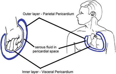
Fig. 5.4
The pericardium. The pericardium is the covering around the heart that is composed of two distinct but continuous layers that are separated from each other by a potential space containing a lubricating serous fluid. During embryological development, the heart migrates into the celomic cavity and a serous lining wraps around it, a process similar to a fist being pushed into a balloon (the balloon and pericardium is one continuous layer of material). The pericardium can be divided into the visceral pericardium (epicardium) and the parietal pericardium. A small amount of serous fluid is secreted into the pericardial space to lubricate the movement of the epicardium on the parietal pericardium. The parietal pericardium contains an epipericardial layer called the fibrous pericardium
Inferiorly, the parietal pericardium is attached to the diaphragm. Anteriorly, the superior and inferior pericardiosternal ligaments secure the parietal pericardium to the manubrium and the xiphoid process, respectively. Laterally, the parietal pericardium (specifically, the fibrous pericardium) is in contact with the parietal pleura (the covering of the lungs). Trapped between the fibrous pericardium and the parietal pleura are the phrenic nerves (motor innervation to the diaphragm). Accompanying these nerves are the pericardiacophrenic arteries and veins (supplying the nerve, pericardium, and diaphragm).
Under normal circumstances, only serous fluid exists between the visceral and parietal layers in the pericardial space or cavity. However, the accumulation of fluid (blood from trauma, inflammatory exudate following infection) in the pericardial space leads to the compression of the heart. This condition, called cardiac tamponade (“heart” + tampon = “plug”), occurs when the excess fluid limits the expansion of the heart (the fibrous pericardium resists stretching) between beats and reduces the ability to pump blood, leading to hypoxia (hypo = “low” + “oxygen”).
Superiorly, the parietal pericardium surrounds the aorta and pulmonary trunk (about 3 cm above their departure from the heart) and is referred to as the arterial reflections or arterial mesocardium; the superior vena cava, inferior vena cava, and pulmonary veins are surrounded by the venous reflections or venous mesocardium. The outer fibrous (epipericardial) layer merges with the outer adventitial layer of the great vessels, which is continuous with the visceral pericardium. The result of this reflection is that the heart hangs “suspended” within the pericardial cavity. For more details on the Pericardium, see Chap. 9.
Within the parietal pericardium, a blind-ended saclike recess called the oblique pericardial sinus is formed from the venous reflections of the inferior vena cava and pulmonary veins (Fig. 5.5). A space called the transverse pericardial sinus is formed between the arterial reflections above and the venous reflections of the superior vena cava and pulmonary veins below. This sinus is important to cardiac surgeons in various procedures when it is important to stop or divert the circulation of blood from the aorta and pulmonary trunk. By passing a surgical clamp or ligature through the transverse sinus and around the great vessels, the tubes of a circulatory bypass machine can be inserted. For more details on cardiopulmonary bypass, see Chap . 33.
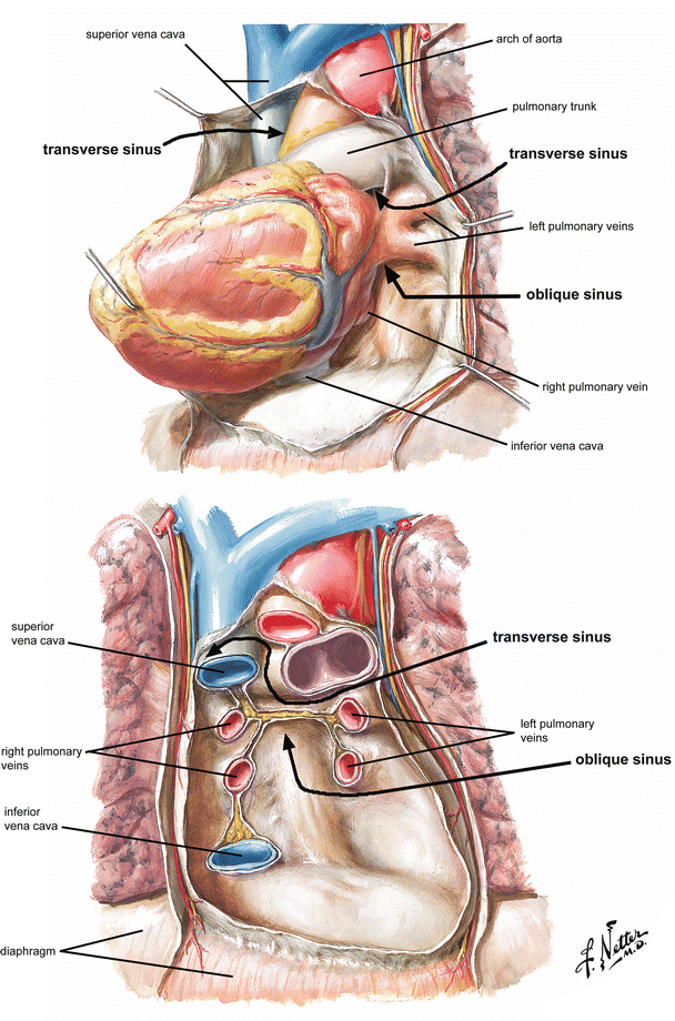
Fig. 5.5
Pericardial sinuses. (a) A blind-ended sac called the oblique pericardial sinus is formed from the venous reflections of the inferior vena cava and pulmonary veins. (b) Another sac, the transverse pericardial sinus, is formed between the arterial reflections above and the venous reflections of the superior vena cava and pulmonary veins below below. © 2006 Elsevier Inc. All rights reserved. www.netterimages.com, Frank Netter
5.4 Internal Anatomy of the Heart
A cross-section cut through the heart reveals a number of layers (Fig. 5.6). From superficial to deep, these are (1) the parietal pericardium with its dense fibrous layer, the fibrous pericardium; (2) the pericardial cavity (containing only serous fluid); (3) a superficial visceral pericardium or epicardium (epi = “upon” + “heart”); (4) a middle myocardium (myo = “muscle” + “heart”); and (5) a deep lining called the endocardium (endo = “within”). The endocardium is the internal lining of the atrial and ventricular chambers and is continuous with the endothelium (lining) of the incoming veins and outgoing arteries. It also covers the surfaces of the AV valves, pulmonary and aortic valves, as well as the chordae tendineae and papillary muscles. The endocardium is a sheet of epithelium called endothelium that rests on a dense connective tissue layer consisting of elastic and collagen fibers. These fibers also extend into the core of the previously mentioned valves.
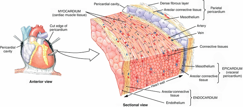
Fig. 5.6
Internal anatomy of the heart. The walls of the heart contain three layers—the superficial epicardium, the middle myocardium composed of cardiac muscle, and the inner endocardium. Note that cardiac muscle cells contain intercalated disks which enable the cells to communicate and allow direct transmission of electrical impulses from one cell to another. Martini, Frederic H.; Timmons, Michael J.; Tallitsch, Robert B., Human Anatomy, 4th, © 2003. Printed and electronically reproduced by permission of Pearson Education, Inc., New York, New York
The myocardium is the tissue of the heart wall, the layer that actually contracts. The myocardium consists of cardiac muscles which are circularly and spirally arranged networks of muscle cells that squeeze blood through the heart in the proper directions (inferiorly through the atria and superiorly through the ventricles). Unlike all other types of muscle cells, (1) cardiac muscle cells branch; (2) cardiac muscles join together at complex junctions called intercalated disks, so that they form cellular networks; and (3) each cell contains single centrally located nuclei. A cardiac muscle cell is not called a fiber. The term cardiac muscle fiber, when used, refers to a long row of joined cardiac muscle cells.
Like skeletal muscle, cardiac muscle cells are triggered to contract by Ca2+ ions flowing into the cell. Cardiac muscle cells are joined by complex junctions called intercalated disks . The disks contain adherens to hold the cells together, and there are gap junctions to allow ions to pass easily between the cells. The free movement of ions between cells allows for the direct transmission of an electrical impulse through an entire network of cardiac muscle cells. This impulse, in turn, signals all the muscle cells to contract at the same time. For more details on the electrical properties of the heart, the reader is referred to Chap . 13.
5.4.1 Cardiopulmonary Circulation
In order to best understand the internal anatomy of the heart, it is desirable to first understand its general function. The heart has two primary functions—collect oxygen-poor blood and pump it to the lungs for the release of carbon dioxide in exchange for oxygen and collect oxygen-rich blood from the lungs and pump it to all tissues in the body to provide oxygen in exchange for carbon dioxide.
The four chambers in the heart can be segregated into the left and the right side, each containing an atrium and a ventricle. The right side is responsible for collecting oxygen-poor blood and pumping it to the lungs. The left side is responsible for collecting oxygen-rich blood from the lungs and pumping it to all tissues in the body. Within each side, the atria are the sites where blood collects and passes through to the ventricles and then they contract to eject the final volumes of blood into the ventricles. The ventricle is much stronger, and it is a site for the pumping of blood out and away from the heart (Figs. 5.7 and 5.8).
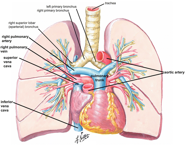
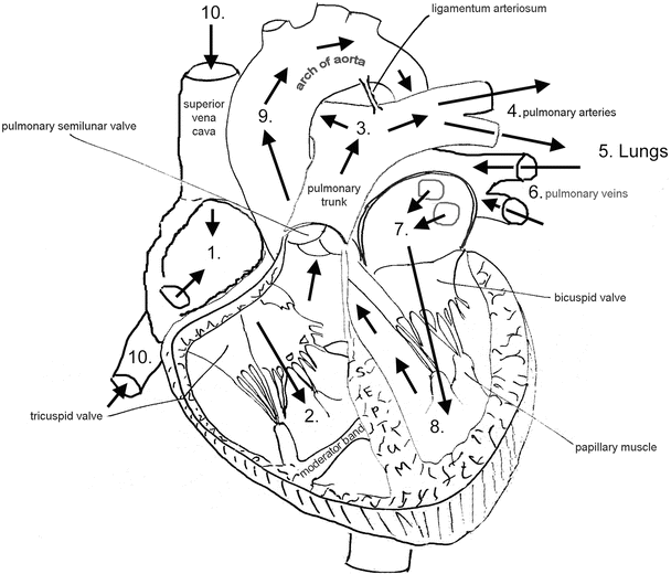
Fig. 5.7
Cardiopulmonary circulation. The four chambers in the heart can be segregated into the left and the right side, each containing an atrium and a ventricle. The right side is responsible for collecting oxygen-poor blood and pumping it to the lungs. The left side is responsible for collecting oxygen-rich blood from the lungs and pumping it to the body. An artery is a vessel that carries blood away from the heart, while a vein is a vessel that carries blood toward the heart. The pulmonary trunk and arteries carry blood to the lungs. Exchange of carbon dioxide for oxygen occurs in the lung through the smallest of vessels, the capillaries. Oxygenated blood is returned to the heart through the pulmonary veins and collected in the left atrium atrium. © 2006 Elsevier Inc. All rights reserved. www.netterimages.com, Frank Netter
Fig. 5.8
Cardiac circulation. Blood collected in the right atrium is pumped into the right ventricle. Upon contraction of the right ventricle, blood passes through the pulmonary trunk and arteries to the lungs. Oxygenated blood returns to the left atrium via pulmonary veins. The left atrium pumps the blood into the left ventricle. Contraction of the left ventricle sends the blood through the aortic artery to all tissues in the body. The release of oxygen in exchange for carbon dioxide occurs through capillaries in the tissues. Return of oxygen-poor blood is through the superior and inferior vena cavae which empty into the right atrium. Note that a unidirectional flow of blood through the heart is accomplished by valves
The right ventricle is the site for the collection of ALL oxygen-poor blood. The large superior and inferior venae cavae, among other veins, carry oxygen-poor blood from the upper and lower parts of the body to the right atrium. The right ventricle pumps the blood out of the heart and through the pulmonary trunk. The term trunk, when referring to a vessel, is a convention that indicates an artery that bifurcates. The pulmonary trunk bifurcates into the left and right pulmonary arteries that enter the lungs. It is important to note that the term “artery” is always used for a vessel that carries blood AWAY from the heart. This is irrespective of the oxygen content of the blood that flows through the vessel.
Once oxygenated, the oxygen-rich blood returns to the heart from the right and left lung through the right and left pulmonary vein, respectively (“vein”—a vessel carrying blood toward the heart). Each pulmonary vein bifurcates before reaching the heart. Thus, there are typically four pulmonary veins entering the left atrium. Oxygen-rich blood is pumped out of the heart by the left ventricle and into the aortic artery.
Observing the heart from a superior vantage point, the pulmonary trunk assumes a leftmost anterior location projecting upward from the base of the heart, the aorta is located in a central location, and the superior vena cava has the rightmost posterior location .
5.4.2 The Right Atrium
The interior of the right atrium has three anatomically distinct regions, each a remnant of embryologic development. The posterior portion of the right atrium has a smooth wall and is referred to as the sinus venarum (embryologically derived from the right horn of the sinus venosus). The wall of the anterior portion of the right atrium is lined by horizontal, parallel ridges of muscle bundles that resemble the teeth of a comb, hence the name pectinate muscle (pectin = “a comb,” embryologically derived from the primitive right atrium). Finally, the interatrial septum is primarily derived from the embryonic septum primum and septum secundum. For more details on the embryology of the heart, refer to Chap. 3.
The smooth posterior wall of the right atrium holds the majority of the named structures of the right atrium. It receives both the superior and inferior vena cavae and the coronary sinus. It also contains the fossa ovalis, the sinoatrial (SA) node, and the AV node.
The inferior border of the right atrium contains the opening or ostium of the inferior vena cava and the os or ostium of the coronary sinus (Fig. 5.9). The coronary sinus is located on the posterior (inferior) side of the heart and receives almost all of the deoxygenated blood from the vasculature of the heart. The os of the coronary sinus opens into the right atrium anteriorly/inferiorly to the orifice of the inferior vena cava. A valve of the inferior vena cava (Eustachian valve, a fetal remnant) guards the orifice of the inferior vena cava (Bartolommeo E. Eustachio, Italian Anatomist, 1520–1574). The valve of the coronary sinus (Thebesian valve) covers the opening of the coronary sinus (fetal remnant to prevent backflow, Adam C. Thebesius, German physician, 1686–1732). Both of these valves vary in size and presence. These two venous valves insert into a prominent ridge, the Eustachian ridge (sinus septum), that runs medial–lateral across the inferior border of the atrium and separates the os of the coronary sinus and inferior vena cava. For more details on the valves of the heart, refer to Chap. 34.
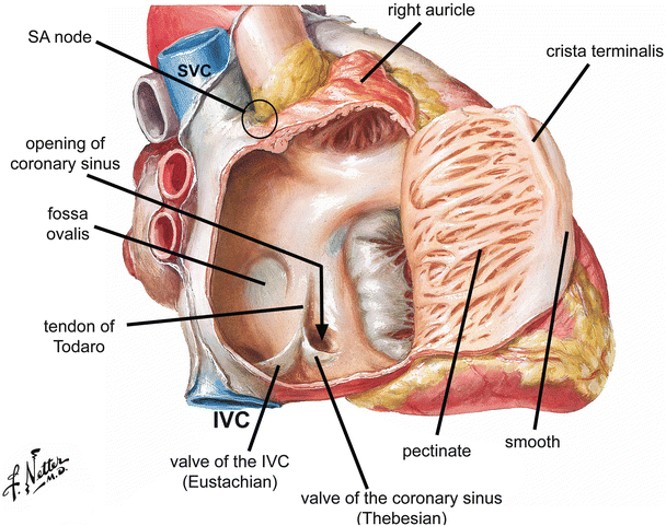
Fig. 5.9
Internal anatomy of the right atrium. The interior of the right atrium has three anatomically distinct regions: (1) the posterior portion (sinus venarum) which has a smooth wall; (2) the wall of the anterior portion which is lined by horizontal, parallel ridges of muscle referred to as pectinate; and (3) the atrial septum septum. © 2006 Elsevier Inc. All rights reserved. www.netterimages.com, Frank Netter
On the medial side of the right atrium, the interatrial septum (atrial septum) has an interatrial and an atrioventricular part. The fossa ovalis (a fetal remnant) is found in the interatrial part of the atrial septum. It appears as a central depression surrounded by a muscular ridge or limbus. The fossa ovalis is positioned anterior and superior to the ostia of both the inferior vena cava and the coronary sinus. A tendinous structure, the tendon of Todaro, crosses the floor of the right atrium. It connects the valve of the inferior vena cava to a portion of the interventricular septum (between ventricles). More specifically, the tendon connects to the central fibrous body (the right fibrous trigone) as a fibrous extension of the membranous portion of the interventricular septum. It courses obliquely within the Eustachian ridge and separates the fossa ovalis above from the coronary sinus below. This tendon likely has a structural role to support the inferior vena cava via the Eustachian valve and is a useful landmark in approximating the location of the AV node (conduction system).
To approximate the location of the AV node, found in the floor of the right atrium and the atrial septum, it is necessary to form a triangle (triangle of Koch; Walter Koch, German surgeon, unknown–1880) using the following structures: (1) the os of the coronary sinus, posteriorly; (2) the right AV opening, anteriorly; and (3) the tendon of Todaro, posteriorly (Fig. 5.10).
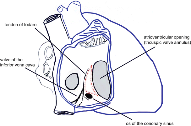
Fig. 5.10
Koch’s triangle. Three landmarks are used to triangulate (dotted red lines) the location of the atrioventricular node (Taware’s node) of the conduction system: (1) coronary sinus, (2) atrioventricular opening, and (3) tendon of Todaro
In the lateral wall and the septum of the smooth portion of the right atrium are numerous small openings in the endocardial surface. These openings are the ostia of the smallest cardiac (Thebesian) veins. These veins function to drain deoxygenated blood from the myocardium to empty into the right atrium which is the collecting site for all deoxygenated blood (for more details on cardiac vasculature, see Chap. 8).
In the anterior–superior portion of the right atrium, the smooth wall of the interior becomes the pectinate portion of the right atrium. The smooth and pectinate regions are separated by a ridge, the crista terminalis (crista = “crest” + “terminal”). The ridge represents the end of the smooth wall and the beginning of the pectinate wall. It begins at the junction of the right auricle with the atrium and passes inferiorly over the “roof” of the atrium. The crista runs inferiorly and parallel to the openings of the superior and inferior vena cavae. As early as the developing embryo, the crista terminalis separates the sinus venosus and the primitive atrium and remains to separate the smooth and the pectinate portion of the right atrium in the definitive heart. The crista terminalis on the internal side results in a groove on the external side of the atrium called the sulcus terminalis.
The SA node is the “pacemaker” of the conduction system. The SA node is located between the myocardium and epicardium in the superior portion of the right atrium. The intersection of three lines indicates the location of the SA node: (1) the sulcus terminalis, (2) the lateral border of the superior vena cava, and (3) the superior border of the right auricle (Fig. 5.11). The name of the SA node is derived from its location between the sinus venarum and primitive atrium. The crista terminalis is the division between these two components in the fetus and adult. It seems logical that the sulcus terminalis is a useful landmark for the approximation of the location of the SA node .
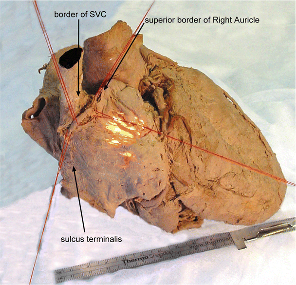
Fig. 5.11
Location of the sinoatrial node. Human cadaver heart demonstrating that the intersection of three lines indicates the position of the sinoatrial node (pacemaker of the conduction system) in the smooth muscle portion of the right atrium: (1) the sulcus terminalis, (2) the lateral border of the superior vena cava, and (3) the superior border of the right auricle. Note the muscle fiber bundles in the wall of the pectinate portion of right atrium. IVC inferior vena cava, SVC superior vena cava
5.4.3 The Right Ventricle
The right ventricle receives blood from the right atrium and pumps it to the lungs through the pulmonary trunk and arteries. Most of the anterior surface of the heart is formed by the right ventricle (Fig. 5.12). Abundant, coarse trabeculae carneae (“beams of meat”) characterize the walls of the right ventricle. Trabeculae carneae are analogous to pectinate muscle of the right atrium and are found in both the right and left ventricles. The outflow tract, conus arteriosus (“arterial cone”), and infundibulum (“funnel”) carry blood out of the ventricle in an anterior–superior direction and can be quite variable in structure—smooth walled or highly trabeculated. A component of the conus arteriosus forms part of the interventricular septum. This small septum, the infundibular (conal) septum, separates the left and right ventricular outflow tracts and is located just inferior to both semilunar valves. Four distinct muscle bundles, collectively known as the semicircular arch , separate the outflow tract from the rest of the right atrium. These muscle bundles are also known as the supraventricular crest and the septomarginal trabeculae .
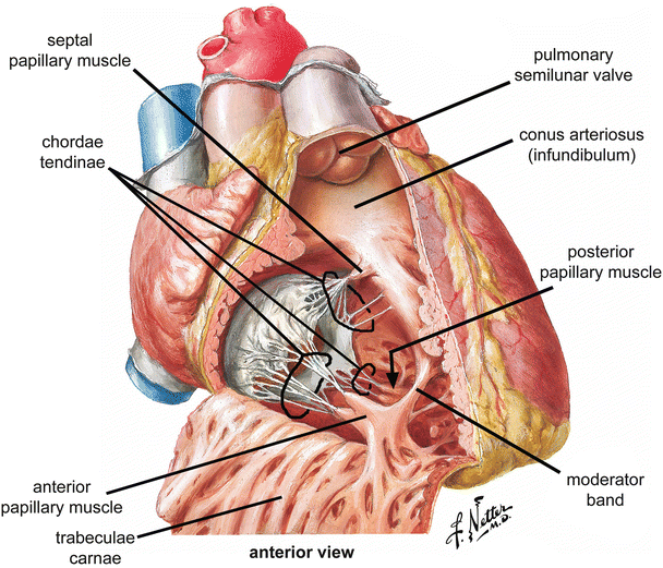
Fig. 5.12
Internal anatomy of the right ventricle. Coarse trabeculae carneae characterize the walls of the right ventricle. The conus arteriosus makes up most of the outflow tract. The right atrioventricular or tricuspid valve is made up of three sets of cusps, chordae tendineae, and papillary muscles. © 2006 Elsevier Inc. All rights reserved. www.netterimages.com, Frank Netter
5.4.3.1 Tricuspid Valve
Blood is pumped from the right atrium through the AV orifice into the right ventricle. When the right ventricle contracts, blood is prevented from flowing back into the atrium by the right AV valve or tricuspid (“three cusps”) valve. The valve consists of the annulus, three valvular leaflets, three papillary muscles, and three sets of chordae tendineae (Figs. 5.12 and 5.13). The AV orifice is reinforced by the annulus fibrosus of the cardiac skeleton (dense connective tissue). Medially, the annulus is attached to the membranous interventricular septum.
 < div class='tao-gold-member'>


Only gold members can continue reading. Log In or Register to continue
Stay updated, free articles. Join our Telegram channel
Full access? Get Clinical Tree
