Chapter 49 Acrocyanosis
The term acrocyanosis is derived from the Greek words akron (meaning “extremity”) and kyanos (meaning “blue”). As a medical condition, acrocyanosis is an uncommon functional vasospastic disorder characterized by persistent bluish discoloration, primarily of the hands and feet. The original description of acrocyanosis is credited to Crocq in 1896, thus the eponym Crocq’s disease. Classically, acrocyanosis affects young asthenic females in regions with cooler climates. In contrast to Raynaud’s syndrome, the bluish discoloration of acrocyanosis is persistent rather than episodic and is not associated with discomfort.1 Primary acrocyanosis is not associated with an identifiable cause and is usually a benign disorder with a good prognosis; skin ulceration and tissue loss are extremely rare. Patients with primary disease usually do well with behavioral measures such as avoidance of exposure to cold; pharmacological therapy is rarely necessary. Secondary acrocyanosis occurs in association with another underlying disorder. An acrocyanotic appearance may be seen in conjunction with any disease that causes central cyanosis or markedly decreases blood flow to the extremity. Treatment and prognosis of secondary acrocyanosis depends on the underlying condition.1
Epidemiology
Limited information is available on many of the epidemiological aspects of primary acrocyanosis, including its precise incidence and prevalence. Acrocyanosis is believed to occur more frequently with a low body mass index (BMI), outdoor occupations, and in areas with cooler climates.2 Despite early reports suggesting similar gender predilection, acrocyanosis seems to affect women more frequently than men, with a female-to-male ratio of 6:1.3 It is more common in younger individuals, the typical age range being 20 to 45 years.4 In addition, occurrence of acrocyanosis seems to decrease with increasing age, regardless of the regional climate.5 It is seen frequently in patients with anorexia nervosa, affecting up to 20% of women with this disorder.6 It has been suggested that as many as 10% of patients have a family history of acrocyanosis in one of the first-degree relatives, suggesting a genetic basis.4
Etiology
As already noted, acrocyanosis is commonly a primary or idiopathic disorder that is not associated with an identifiable cause. It also occurs as a secondary condition in association with connective tissue disorders, some hematological conditions, anorexia nervosa, neurovascular disorders, drugs, toxins, infections, heritable metabolic diseases, and some malignancies.7 An acrocyanotic appearance also occurs in conjunction with any disease that causes central cyanosis or markedly decreases blood flow to the extremities (Box 49-1).
Pathophysiology
Acrocyanosis is due to a decrease in the amount of oxygen delivered to the tissues of the extremities. Several mechanisms for primary acrocyanosis have been proposed, but the precise mechanism remains elusive. Potential pathophysiological disturbances include abnormal arteriolar tone, alteration of microvascular responsiveness with capillary and venular dilation and stasis, and abnormal sympathetic nervous system activity (Box 49-2).
The essential abnormality in primary acrocyanosis seems to be peripheral cutaneous vasoconstriction due to increased tone of the arterioles, associated with secondary vasodilation of capillaries and subpapillary venous plexi.8 Arteriolar vasoconstriction produces the cyanotic discoloration, and compensatory venular dilation in the postcapillary sphincter leads to excessive sweating. Persistent vasoconstriction at the precapillary sphincter creates a local hypoxic environment that may cause increased release of vasodilatory mediators such as adenosine in the capillary beds.9 This in turn leads to dilation of postcapillary venules. The difference in the vessel tone may create a countercurrent exchange system in an attempt to retain heat, and this leads to sweating. Potential mediators that may contribute to the pathogenesis of acrocyanosis include serotonin, adrenaline, noradrenaline, and endothelin (ET)-1. Levels of these mediators have been shown to be increased in acrocyanosis.3,10
The exact cause of the disordered arteriolar tone in acrocyanosis is unknown. Most evidence points to local sensitivity of the arterioles to cold and an exaggerated sympathetic nervous system response. Earlier studies suggested an exaggerated digital arteriolar vasoconstrictive response to cold stimuli,11 whereas subsequent studies have implicated the sympathetic nervous system.1 An interesting case described normalization of cutaneous thermal-induced vasoreactivity in the hands of a young girl with acrocyanosis during phenobarbital-induced sleep. In this state, her hands responded in the same manner as the rest of body to heat and cold exposure. This finding supports the hypothesis that the abnormal vascular tone in acrocyanosis may be of central origin.12 Other mechanisms hypothesized include increased blood viscosity,13 decreased distal blood flow at low temperatures, and persistently elevated vasoconstrictive mediators such as endothelin-1 levels,14 but convincing supporting evidence for these is not readily available.
Clinical Presentation
Primary acrocyanosis typically occurs in thin young women who are not physically active. They commonly present with persistent bluish discoloration of the hands and feet. The distribution of the discoloration is symmetrical.5 It less frequently involves forearms, ears, lips, nose, or nipples. It is the discoloration that usually prompts patients to seek medical care. Patients may also describe a sensation of coolness of the affected areas, but generally will not have pain. There may be slight swelling of the digits and increased perspiration. Although the discoloration is persistent, emotion and cold temperature can intensify the acrocyanotic appearance, and warmth can diminish it.1 Discoloration and coolness are worse in cold weather, but typically persist even during the summer months.
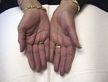
Examination reveals bluish discoloration of the hands and feet, which are cool and often sweaty (Fig. 49-1). The color has also been described as bluish-pink or orange-like tinged with red, and differing hues may be seen at various times. A brownish-yellow color on the dorsum of hands and feet has been described and attributed to exposure to the warmth of the sun because it is most obvious in summer. Pressure on the discolored skin causes pallor, and color returns slowly and irregularly. Fingers may be puffy, but trophic changes do not occur.
< div class='tao-gold-member'>
Stay updated, free articles. Join our Telegram channel

Full access? Get Clinical Tree


