Cells termed pacemakers (see Chapter 1)—located in the sinoatrial (SA) node, at various sites in the atria, and throughout the His–Purkinje network—have the capacity for spontaneous depolarization. Atrial and ventricular muscle cells and atrioventricular (AV) nodal cells do not have pacemaking capabilities. The rate of impulse formation by pacemaker cells is determined by the rate of their spontaneous depolarization, and the more superior the location of the pacemaking cell, the more rapidly this depolarization occurs. Accelerated automaticity is considered to be a tachyarrhythmia only when the heart rate exceeds the arbitrary limit of 100 beats per minute. Because the upper limit of normal automaticity of the SA nodal and atrial cells is 100 beats per minute, any acceleration in the rate of automaticity of these cells is considered a tachyarrhythmia. The upper limit of normal automaticity is 60 beats per minute in the common bundle and 50 beats per minute in the bundle branches. The rhythm produced is called an accelerated rhythm until it reaches 100 beats per minute (Table 16.1).

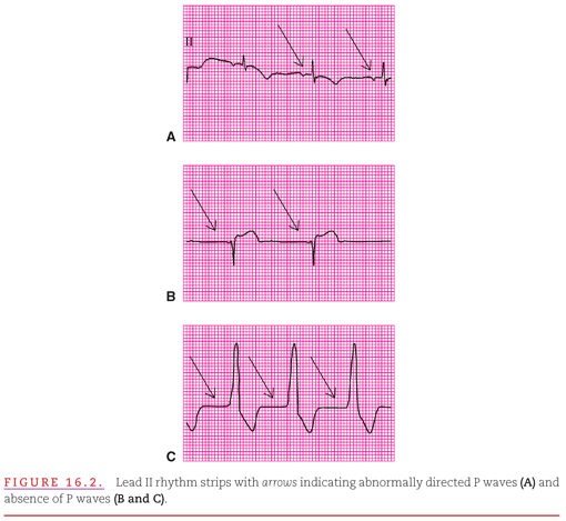
Examples of arrhythmias due to accelerated atrial, junctional, and ventricular automaticity are presented in Figure 16.2. In Figure 16.2A, the accelerated atrial rhythm (AAR) is apparent from the frequent, regular (evenly spaced), but “different from sinus” P waves. In Figure 16.2B, the accelerated junctional rhythm (AJR) is apparent from the frequent, regular, narrow QRS complexes not preceded by P waves. In Figure 16.2C, the accelerated ventricular rhythm (AVR) is apparent from the frequent, regular, wide QRS complexes not preceded by P waves.

Usually, the cardiac rhythm is controlled by the SA node. However, there are several reasons why the rhythm becomes dominated by the accelerated pacing activity from a nonsinus site. These include:
1. A pharmacologic agent that selectively increases automaticity in lower pacemakers.
2. Blockage of sinus impulses in the AV conduction system, permitting an escape focus in the common bundle to control the ventricular rhythm.
3. Local pathology (especially ischemia) that induces automaticity in lower areas with pacemaking capability.
4. Local pathology that decreases automaticity within the SA node.
The rate of cardiac impulse formation is regulated by the balance between the parasympathetic and sympathetic divisions of the autonomic (or involuntary) nervous system. The more superior the location of pacemaking cells, the greater is the degree of their autonomic regulation. An increase in parasympathetic activity decreases the rate of impulse formation, whereas an increase in sympathetic activity increases the rate. The sympathetic nervous system becomes activated by any condition that requires “flight or fight.” Sinus tachycardia that results is therefore a physiologic response to the body’s needs rather than a pathologic cardiac condition. By this principle, treatment of the tachyarrhythmia should be directed at correcting the underlying condition and not at suppression of the SA node itself. Maximal sympathetic stimulation can increase the heart rate produced by the SA node to 200 beats per minute or, rarely, 220 beats per minute in younger individuals. The generally accepted formula for maximal sinus rate is 220 beats per minute subtracted by age. The rate rarely exceeds 160 beats per minute in nonexercising adults.
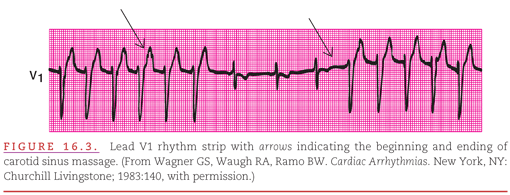
In sinus tachycardia, there is normally one normal P wave for every QRS complex, but coexisting abnormalities of AV conduction may alter this relationship. The PR interval is shorter than during normal sinus rhythm because the increased sympathetic tone that produces the sinus tachycardia also speeds AV nodal conduction. The QRS complex is usually normal in appearance but can be abnormal either because of a fixed intraventricular conduction disturbance (such as bundle-branch block, hypertrophy, or myocardial infarction) or because the rapid rate of activation does not permit time for full recovery of the intraventricular conduction system before the arrival of the next impulse (see Chapter 6). Figure 16.3 demonstrates sinus tachycardia with LBBB. Conduction block is proven to be due to rate-related aberrancy when it disappears during carotid sinus massage–induced sinus slowing or AV block and returns when the rate gradually increases following the massage.

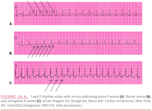
Although the other tachyarrhythmias caused by accelerated automaticity and discussed in this chapter do not mimic sinus tachycardia, a common clinical problem is the differentiation of sinus tachycardia from various reentrant tachyarrhythmias that are discussed in later chapters. If discrete P waves (with anterograde orientation), a short PR interval, and a normal QRS complex duration are present, the diagnosis of sinus tachycardia is most likely. True sinus tachycardia is shown in Figure 16.4A, and two reentrant supraventricular tachyarrhythmias that have a similar appearance to sinus tachycardia are shown in Figure 16.4B and C . When apparent sinus tachycardia is associated with a prolonged PR interval, one should suspect that it is not really a sinus tachycardia. Figure 16.4B presents an example of the reentrant atrial tachyarrhythmia of typical sawtooth-like atrial flutter (see Chapter 17) with only every other flutter wave conducted to the ventricles (2:1 AV conduction). An abnormal limb lead P-wave axis also suggests a nonsinus origin of the tachyarrhythmia. Figure 16.4C presents an example of reentrant junctional tachycardia (see Chapter 18) with inverted P waves following each QRS complex and long PR interval.

When the appearance of atrial activity fails to provide the foregoing clinical differentiation of the source of a tachyarrhythmia, it may be necessary to observe the onset and termination of the tachyarrhythmia: A gradual change in rate establishes the diagnosis of sinus tachycardia (Fig. 16.5A
Stay updated, free articles. Join our Telegram channel

Full access? Get Clinical Tree


