MAGNETIC RESONANCE IMAGING OF THE CARDIAC SCAR
Case presented by:
A.Contrast-enhanced MR angiography (CEMRA) for visualizing three-dimensional (3D) left atrial and pulmonary vein (PV) anatomy.
B.3D delayed late gadolinium enhancement (LGE) for visualizing postablation scars in the left atrium.
C.Replacing electrical mapping of the left atrium.
D.A and B.
E.A and C.
Discussion
CEMRA is a 3D spoiled gradient echocardiography-based technique that can be used with or without cardiac gating to visualize the vascular structures such as left atrium, PVs, or aorta. Cardiac gating is shown to improve cardiac motion suppression1 and will result in sharper delineation of the PVs and left atrial border. Postprocessing software is often used to segment the left atrium from the rest of the nearby cardiac structures. The high-resolution left atrial 3D model can be reviewed preablation as a guiding roadmap, or integrated into an EP electroanatomic mapping system2 during the ablation procedure for optimal accuracy in localization (Figure 74.1). A recent study showed that the integration of an EP electroanatomic mapping system with 3D CEMRA data could reduce fluoroscopy time, although it did not improve overall patient outcome3.
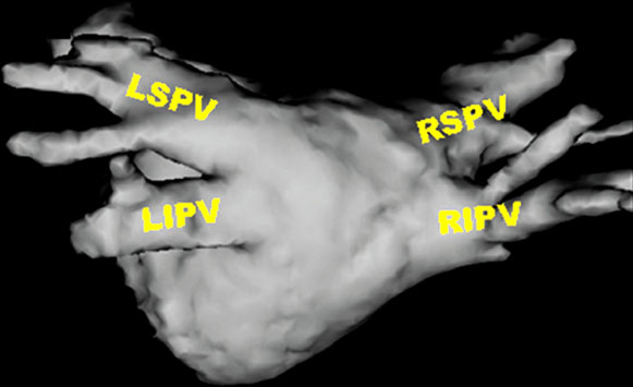
Figure 74.1. A left atrial 3D volume rendering of a MR angiogram. LIPV, left inferior pulmonary vein; LSPV, left superior pulmonary vein; RIPV, right inferior pulmonary vein; RSPV, right superior pulmonary vein.
MR delayed LGE has been an established method to help assess myocardial scarring in various heart diseases such as myocardial infarction and hypertrophic cardiomyopathies. The mechanism of LGE relies on the expanded extracellular space in fibrotic tissue and the slower perfusion of infarcted tissue. Since the infarcted/fibrotic tissue has a longer washout period post injection, the contrast media stays within these tissues longer than normal tissue. Therefore, the infarcted area will have shorter T1 (ie, elevated signal intensity in LGE) when imaged 10 to 20 min post injection. The LGE application employs an inversion recovery pulse to invert the Mz magnetization of all the tissue, and then images with a T1-weighted fast gradient echo sequence after a certain inversion (TI) time. The TI time is selected such that short T1 species (eg, scar) will recover faster and have positive signal, whereas longer T1 species (eg, normal tissue) will be at null point (ie, zero Mz magnetization).
Delayed Enhancement in Ventricle
Stay updated, free articles. Join our Telegram channel

Full access? Get Clinical Tree


