CT IMAGING OF THE LEFT ATRIUM
Case presented by:
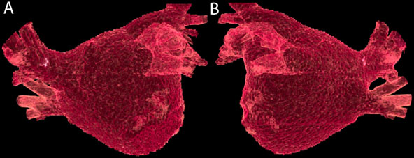
Figure 73.1. Anterior (A) and posterior (B) views of a segmented left atrial model from computed tomography (CT) scanning.
Question No. 1: The images in Figure 73.1:
A.Show the left atrium (LA) of a patient with atrial fibrillation (AF) who has pulmonary vein (PV) narrowing.
B.Depict the anatomy from a patient with a very common two-right-by-two-left PV anatomy and are useful in preparing a road map for ablation.
C.Were obtained with the use of contrast.
D.B and C above.
Discussion
Three-dimensional (3D) imaging of the heart in patients undergoing left atrial procedures is becoming popular because it accurately depicts the anatomy of the LA and PVs.1 This information can assist in preprocedure planning, and the resultant segmented images may be integrated into a cardiac mapping system to aid in the ablation.2,3 Cardiac CT has the advantage of better spatial resolution when compared to magnetic resonance imaging (MRI).
Compensation must be made for motion of the heart due to the respiratory and cardiac cycles to obtain good image quality. The CT scan is acquired quickly enough that it can be performed during a single breath hold for most patients. Cardiac cycle motion can be “frozen” through the use of ECG gating. During the scan, wherein image data arebeing acquired continuously, the patient’s surface ECG is also being recorded. Then, a series of two-dimensional (2D) axial images (Figure 73.2) are retrospectively recreated from only the raw scan data that correspond to a particular point in the cardiac cycle. The images, which are acquired over several rotations of the gantry around the patient, will then correspond to one another, essentially eliminating motion of the heart from the series.
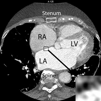
Figure 73.2. 2D axial slice from CT scanning showing the right atrium (RA), left atrium (LA) and left ventricle (LV), as well as the spine and sternum. The insert shows the individual pixels that make up the 2D image.
Stay updated, free articles. Join our Telegram channel

Full access? Get Clinical Tree


