INTERVENTIONAL TECHNIQUES TO SUCCESSFULLY ADD RV PACE-SENSE AND LV LEADS DESPITE MULTIPLE PITFALLS
Case presented by:
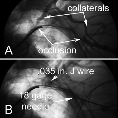
The following 12 Figures were taken from a single implant procedure in a patient with a Sprint Fidelis® lead (Medtronic Inc., Minneapolis, MN) who had a left bundle branch block (LBBB), class III congestive heart failure (CHF), and an ejection fraction of 27%. She had the addition of a left ventricular (LV) lead and a right ventricular (RV) pace-sense lead. To reduce the risk of nephropathy with the use of contrast, she was hydrated with normal saline 3 mL/kg for 1 hour before the procedure, 1 mL/kg per hour during the procedure, and 1 mL/kg for 6 hours after the procedure.1 At the time of implant, a venogram was obtained via contrast injection through a peripheral vein in the left arm. The venogram (Figure 63.1A) demonstrates an occlusion of the axillary/subclavian vein with extensive collaterals.
Question No. 1: Based on the venogram, which of the following is true?
A.Access is not possible from the left; move to the right.
B.Access is not possible; extract one of the leads to gain access on the left.
C.Despite the apparent total occlusion, it may be possible to gain access on the left.
Figure 63.1. Insertion of a J-wire despite apparent long total occlusion on peripheral vein venogram. In Panel A, contrast injected through a peripheral vein demonstrates an apparent long total occlusion with extensive collaterals. In Panel B, the needle is inserted into the axillary vein at the site of apparent occlusion. The 0.035-inch J-wire advances through the site of apparent occlusion but will not advance beyond the clavicle into the subclavian vein. The peripheral vein venogram does not define the location or extent of occlusion.
Figure 63.1B illustrates that the location of the occlusion was not where it appeared on the venogram. The peripheral venogram usually overestimates the severity and location of the occlusion associated with chronic leads.2
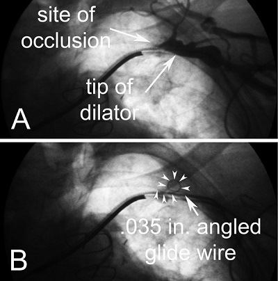
The venogram in Figure 63.2A was then obtained by injection of contrast through the dilator from a 5-Fr sheath advanced as far as possible over the J-wire.
Figure 63.2. The angled 0.035-inch hydrophilic (glide) wire (Angled Glidewire [GR3506], Terumo Medical Corp., Somerset, NJ, or Angled Zipwire [46-151B], Boston Scientific Corp., Natick, MA) will not advance through the dilator from a 5-Fr sheath beyond the site of true obstruction. In Panel A, the dilator from a 5-Fr sheath advanced over the J-wire to the site of obstruction and contrast injected. There is no apparent opening in the occlusion when contrast is injected through the dilator. In Panel B, the glide wire (arrowheads) curls when advanced through the dilator and will not proceed beyond the obstruction.
Question No. 2: Based on the local venogram, which of the following is true?
A.Access is not possible from the left; move to the right.
B.Access is not possible; extract one of the leads to gain access on the left.
C.Despite the apparent total occlusion, it may be possible to gain access on the left.
Despite the apparent total occlusion, an attempt was made to advance a glide wire through the occlusion but was unsuccessful (see Figure 63.2B).
Question No. 3: Based on the failure of the wire to advance, which of the following is indicated?
A.Access is not possible from the left; move to the right.
B.Access is not possible; extract one of the leads to gain access on the left.
C.Despite the failure of the glide wire to advance, it still may be possible to gain access on the left.
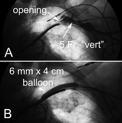
The venogram in Figure 63.3A was obtained by contract injection through a left vertebral diagnostic catheter and demonstrated an opening in the obstruction. The glide wire is advanced using the direction and support of the catheter, and venoplasty was performed to gain access.3 Because the patient had a Sprint Fidelis defibrillator lead, an attempt was made to place an RV pace-sense lead in the setting of severe tricuspid regurgitation and massively enlarged right atrium. The lead repeatedly dislodged despite using stiff stylets and replacing the lead.
Figure 63.3. An opening is identified by injection of contrast through a 5-Fr left vertebral diagnostic catheter (“vert”), the glide wire advanced, and venoplasty performed. In Panel A, contrast injection through the “vert” catheter (Vert Slip-Cath Beacon® Tip [SCBR5.0-38-100-P-NS-VERT], Cook Medical Inc., Bloomington, IN, or Angled Taper Terumo Glidecath® [CG508], Terumo Medical Corp.) reveals an opening in the occlusion. Before inserting the “vert,” the dilator is exchanged for a 5-Fr sheath. The tip of the “vert” is directed toward the leads where contrast injection defines the opening. The “vert” catheter provides direction and support to advance the glide wire through the opening. In Panel B, the obstruction is dilated with a 6-mm diameter by 4-cm length balloon, inflated to rated burst pressure (14 atmospheres). With the glide wire in the pulmonary artery or the inferior vena cava, the “vert” catheter is advanced to the tip of the glide wire, and the glide wire exchanged for a 0.035-inch extra support wire (Amplatz Extra-Stiff Wire Guide, 0.035-inch, 145-cm, 3-mm tip curve [THSCF-35-145-3-AES], Cook Medical Inc.).
Question No. 4: Based on the difficulty in placing the RV lead, which of the following is indicated?
A.Keep trying with stiff stylets; the lead will eventually stay put.
B.Give up and continue to use the pace-sense portion of the Sprint Fidelis lead.
C.Use a long anatomically shaped sheath to provide support for the lead as it is screwed in place.
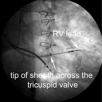
Figure 63.4 illustrates the use of a long anatomically shaped sheath to secure an RV lead despite severe tricuspid regurgitation and massively enlarged right atrium. Following placement of the RV lead, attempts were made to cannulate the coronary sinus (CS). The CS could not be located after 20 minutes, despite using an electrophysiology (EP) catheter and probing wire.
Figure 63.4. Long anatomically shaped sheath (SafeSheath® CSG® Worley Braided Core Series 40-cm long, 9-Fr I.D. [CSG/Worley/BCore-1-09], Pressure Products Inc., San Pedro, CA) providing support to secure RV lead despite severe tricuspid regurgitation.
Stay updated, free articles. Join our Telegram channel

Full access? Get Clinical Tree


