BUNDLE BRANCH REENTRY VENTRICULAR TACHYCARDIA
Case presented by:
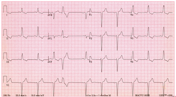
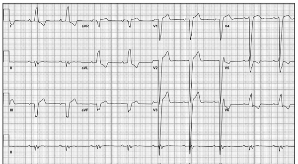
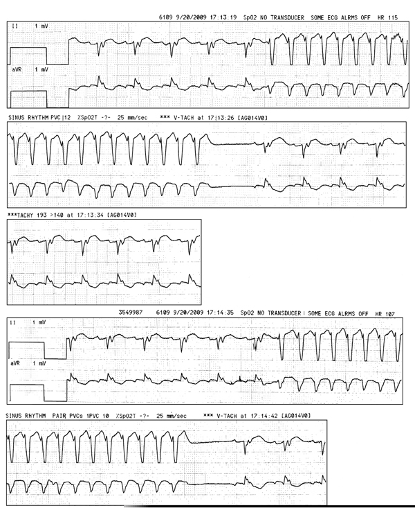
A 57-year-old man underwent mechanical aortic valve replacement for severe aortic insufficiency due to a bicuspid valve. Preoperative evaluation revealed a severely dilated left ventricle (LV) (6.9 cm at end diastole and 5.8 cm at end systole), left ventricular ejection fraction (LVEF) 54% and no coronary artery disease by coronary angiography. The ECG is shown in Figure 38.1. Three days after discharge, he had 2 syncopal episodes at home. Echocardiogram showed normal prosthetic valve function and LVEF of 49%; ECG is shown in Figure 38.2. Inpatient cardiac monitoring for 36 hours revealed sinus rhythm and no significant arrhythmias. On postoperative day 3, he had developed runs of self-terminating wide-complex tachycardia at 250 bpm (Figure 38.3). He was treated with amiodarone for the same.
Question No. 1: What is the likely cause of this patient’s syncopal spells?
A.Vasovagal syncope.
B.Intermittent heart block.
C.Rapid atrial arrhythmia.
D.Monomorphic ventricular tachycardia (VT).
E.Bundle branch reentry (BBR) VT.
Question No. 2: What would be the most appropriate management?
A.Increase amiodarone and metaprolol doses.
B.Outpatient long-term cardiac monitoring.
C.Transesophageal echocardiography to assess prosthetic valve function.
D.Electrophysiologic studies.
E.Dual-chamber implantable cardioverter-defibrillator (ICD) implantation.


Stay updated, free articles. Join our Telegram channel

Full access? Get Clinical Tree


