ECG LOCALIZATION OF ACCESSORY PATHWAYS
Case presented by:
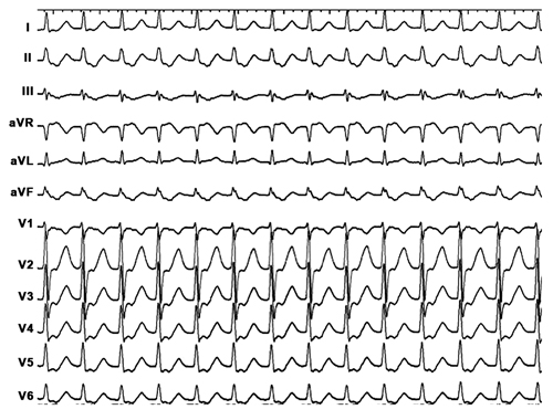
A 49-year-old male presents with long-standing history of palpitations and a recent syncopal episode. Echocardiogram did not show any structural heart disease. The baseline ECG is shown in Figure 13.1.
Figure 13.1 Baseline ECG.
Question No. 1: Your diagnosis is ventricular preexcitation over a:
A.Right free wall accessory pathway (AP).
B.Right septal AP.
C.Left posterior AP.
D.Left lateral AP.
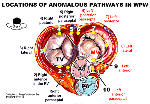
Figure 13.2. Location of various APs in patients with Wolff-Parkinson-White (WPW) syndrome. (Reproduced from Gallagher et al1 with permission from Elsevier.)
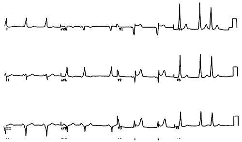
Figure 13.3. 12-lead ECGs of patients with WPW.
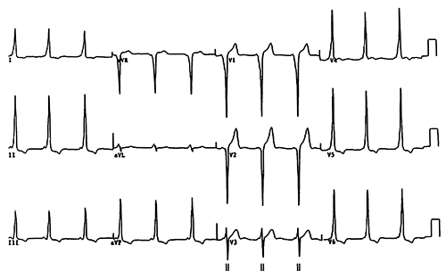
Figure 13.4. 12-lead ECGs of patients with WPW.
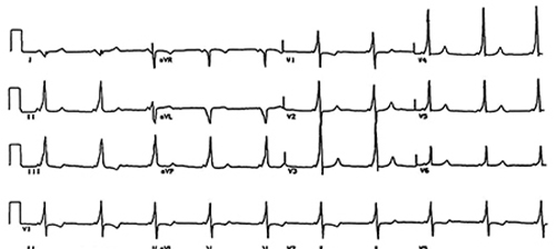
Figure 13.5. 12-lead ECGs of patients with WPW.
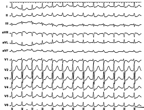
Figure 13.6. 12-lead ECG with narrow QRS complex supraventricular tachycardia (SVT) that was induced in the laboratory to the patient.
Question No. 2: The most likely diagnosis is:
A.Atrial tachycardia (AT) arising from the superior crista terminalis.
B.Typical atrioventricular nodal reentrant tachycardia (AVNRT).
C.Atrioventricular reentrant tachycardia (AVRT) using left posterior AP.
D.AVRT using a right anterior AP.
Figure 13.7. An example showing a 12-lead ECG of another SVT that was initiated in the laboratory in this patient.
Question No. 3: The most likely diagnosis is:
A.AT arising from the coronary sinus (CS).
B.Typical AVNRT.
C.AVRT using left posterior AP.
D.AVRT using a right anterior AP.
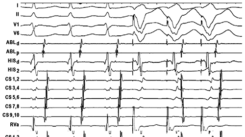
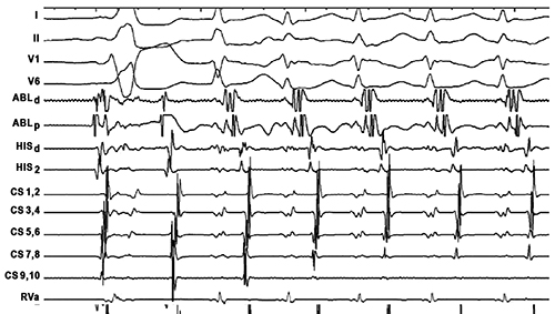
Figure 13.8. Surface ECGs together with recordings from the ablation catheter (Abl) in the high right atrium (HRA), the His bundle (His D, P, and 2), the proximal CS 9,10 to distal CS 1,2, and from the right ventricle (RVa).
Question No. 4: This finding would rule out which one of the following:
A.Atrial tachycardia.
B.Atypical AVNRT.
C.AVRT using a septal AP.
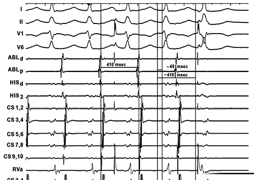
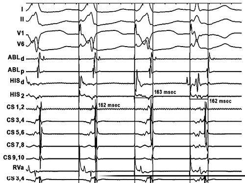
Figure 13.9. Delivery of single premature ventricular impulse during tachycardia.
Question No. 5: This observation proves:
A.AP pathway is present in this patient.
B.AP is integral to the tachycardia hence proving a diagnosis of AVRT.
C.AVNRT cannot be excluded.
D.AT cannot be excluded.
Figure 13.10. An example of para-Hisian pacing.
Question No. 6: Findings shown in Figure 13.10 demonstrate:
A.Retrograde conduction over AV node only.
B.Retrograde conduction over septal AP only.
C.Retrograde conduction over AV node and AP.
D.Absence of retrograde conduction.
Figure 13.11. An example showing surface ECGs together with recordings from the Abl in the right septal region, the His bundle (His D, P, and 2), the proximal CS 9,10 to distal CS 1,2 and from the RVa.
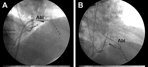
Question No. 7: The ablation site shown in Figure 13.12:
A.Is an ideal site for ablation.
B.Can lead to AV block.
C.Will not affect conduction over septal AP.
Figure 13.12. Ablation site shown in 2 views.
Discussion
Stay updated, free articles. Join our Telegram channel

Full access? Get Clinical Tree


