ATRIAL TACHYCARDIA FROM ABOVE THE SEMILUNAR VALVES
Case presented by:
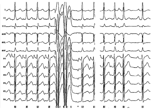
Figure 12.1. ECG of a patient with ectopic atrial tachycardia (AT).
Question No. 1: In the patient with ectopic AT shown in Figure 12.1, the most likely focus is on:
A.Noncoronary sinus.
B.Left pulmonary vein (PV).
C.Superior mitral annulus (SMA).
D.Coronary sinus (CS) ostium.
E.Atrial septum.
Discussion
When P waves during a tachycardia merge with the T wave, introducing premature ventricular complexes brings out the P waves better, as in this case . Morphology of P waves helps focus the mapping efforts to the area of interest.
Stay updated, free articles. Join our Telegram channel

Full access? Get Clinical Tree


