Post-intubation tracheal stenosis may occur at various points in the airway, from the point where an orotracheal tube passes through the vocal cords to the contact point between the tube tip and the distal trachea.
The prolonged presence of an orotracheal tube passing through the vocal cords causes mucosal irritation, which can lead to edema, ulceration, granulation tissue formation and ultimately subglottic stenosis. Tracheostomy generally prevents this complication, although in kyphotic patients pressure on the anterior cricoid may still occur. Laryngeal stenosis in patients with tracheostomy may occur due to infection. Smaller-sized tubes should be used to avoid excessive pressure on the larynx and subglottis.
Formation of bulky granulation tissue at the site of a stoma may occur unrecognized while a tracheostomy tube is in place and lead to airway obstruction immediately after tube removal. Later, the defect through which the tube was placed heals by scarring and contraction. This process converts the cross section of the trachea at the site of the stoma from a C to an A shape with resultant narrowing. This phenomenon is magnified by the presence of an excessively large stoma, either from technical error at the time of surgery or excessive traction from the tube.
Cuff stenosis is the most common complication of prolonged intubation. Pressure necrosis of the tracheal mucosa leads to ulceration and tracheitis and ultimately to erosion of the underlying cartilaginous rings and dense fibrosis of the airway. Prior to the introduction of low-pressure cuffs, the incidence of symptomatic tracheal stenosis was as high as 20% following prolonged intubation. Prolonged high cuff pressure may also lead to tracheoesophageal fistula (described later) or tracheo-innominate fistula.
Tracheal narrowing may occur due to granulation tissue deposition at the site of contact between the tip of the endotracheal tube and the trachea. This complication is less common with modern, concentrically expanding cuff balloons because contact between the tube tip and tracheal wall is limited.
Because many survivors of prolonged intubation have complex medical problems, conservative management of post-intubation stenosis is often initially favored. Repetitive dilatation or laser ablation may be successful in some patients. Airway patency can be maintained indefinitely by creating a new tracheostomy and using a T-tube to splint the airway open. The sidearm of the T-tube is kept closed and used to access the trachea for cleaning and for periodic T-tube changes as necessary. In placing a T-tube, the stoma should be created through the most damaged part of the trachea to preserve viable trachea for future reconstruction.
Carefully selected patients can be managed by tracheal resection and reconstruction (described later) with a success rate exceeding 95%. Expandable stents are generally contraindicated in benign stenosis. Stents can lead to the deposition of granulation tissue proximal and distal to the stent, potentially converting a short, resectable lesion into a long, unresectable one.
Traumatic tracheal stenosis
Blunt injury to the trachea, carina or bronchi may initially go unrecognized. There is usually a history of pneumothorax, frequently bilateral, requiring tube thoracostomy. Tracheostomy below the level of injury may have been performed. Over time the injured segment of trachea becomes stenotic, and if a tracheostomy is present, the upper airway may become totally occluded. Treatment is by surgical repair several months after injury when local inflammation has subsided.
Stenosis due to inhalation injury
Chemical or thermal injury due to the upper airway is challenging to manage. Injury is usually confined to the subglottic and tracheal mucosa, sparing the tracheal rings. Whereas the pharynx and supraglottic larynx often heal without functional deficit, the subglottic airway may develop fibrosis that is difficult to distinguish from intubation-related stenosis. Protecting the airway carefully with silicone T-tubes permits resection and reconstruction, if needed, to be deferred until inflammation has resolved – often many months later.
Post-therapeutic
Recurrent tracheal stenosis may be a late complication of tracheal resection and reconstruction. This can be caused by interruption of the blood supply, excessive anastomotic tension, granulation tissue or recurrence of primary disease. Reconstruction following radiotherapy should be approached with great caution but is feasible in highly selected patients. Careful assessment of the tracheal architecture is essential. The anastomosis should be covered with well-vascularized tissue – either muscle or omentum – to buttress and aid healing.
Stenosis due to infection
Tuberculous infection of the upper airways may lead to ulcerative tracheitis that progresses to long-segment circumferential fibrosis. Surgical treatment is complicated by the length of the lesion and the presence of ongoing inflammation. Complete eradication of infecting organisms and repeated dilation is the usual treatment. Infection with Histoplama capsulatum can lead to fibrotic replacement of mediastinal structures, including the trachea and bronchial tree. Enlarged, fibrotic mediastinal lymph nodes due to histoplasmosis may lead to extrinsic airway compression.
Other causes of tracheal stenosis
Relapsing polychondritis is a systemic condition in which cartilaginous structures become inflamed and fibrotic. When the cartilaginous rings of the trachea are affected, the result is progressive stenosis. Wegener’s granulomatosis, sarcoidosis and amyloidosis may also affect the trachea. Because of the diffuse, systemic nature of these diseases, surgical resection and reconstruction are is frequently impossible, and palliative management with a silicone T-tube may be preferable.
Idiopathic tracheal stenosis
Stenosis of the trachea may occur without an antecedent history of trauma, intubation or inflammatory disease. The majority of affected patients are women in the third to fifth decades of life. Symptoms may develop rapidly over a few months or insidiously over years. Most lesions of idiopathic tracheal stenosis are short (1–3 cm), circumferential and centred at the junction between cricoid cartilage and trachea. The subglottic location of these lesions frequently mandates complex laryngotracheal resection (described later). High lesions may require resurfacing of the posterior cricoid plate with a membranous wall tracheal flap.
Intraluminal obstruction (tracheal masses)
Primary neoplasms of the trachea are rare, comprising 1% of bronchial tumours and 2% of upper respiratory tract tumours. In adults, malignant tumours are significantly more common that benign lesions, whereas in children the reverse is true.
Malignant tracheal tumours
Malignant tumours of the trachea are roughly evenly divided between squamous cell carcinoma and adenoid cystic carcinoma. Other primary malignant masses have been described including carcinoid tumours, adenocarcinoma, malignant fibrous histiocytomas and various sarcomas. Locally invasive extrinsic cancers (thyroid, esophagus, larynx, lung, lymphoma) may also lead to tracheal obstruction.
Squamous cell carcinoma
Squamous cell carcinoma of the trachea is primarily a disease of older male tobacco users. Synchronous primary cancers of the larynx or lung are common. Lesions are typically exophytic or ulcerating and occur in the distal third of the trachea. Local invasion and cervical lymph node metastasis at the time of presentation are common. Staging of regional lymph nodes and careful determination of the length of tumour extension are is important prior to attempts at resection and intraoperatively prior to reconstruction. Post-operative radiation therapy is recommended in almost all patients to improve local control. Survival of up to 47% at 5 years and 36% at 10 years has been reported (Figure 6.2).
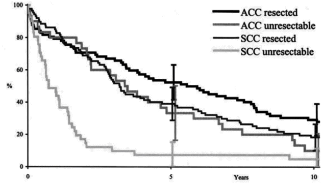
Long-term survival after operative and non-operative treatment of adenoid cystic carcinoma (ACC) and squamous cell carcinoma (SCC) of the trachea.
Adenoid cystic carcinoma
Adenoid cystic carcinoma is not linked to cigarette smoking and has no age predilection. It is more likely to occur in the proximal third of the trachea than squamous cell carcinomas and is slower growing. Compression of adjacent structures in the mediastinum may occur, but true invasion is uncommon. Although lymphatic spread is rare, hematogenous metastases to the lung are frequently seen. A complete surgical resection is preferable, but because of the relatively indolent nature of adenoid cystic carcinoma, a positive resection margin is preferable to an anastomosis under tension. All patients should receive post-operative radiation therapy. Survival of 73% at 5 years and 57% at 10 years is described (Figure 6.3).
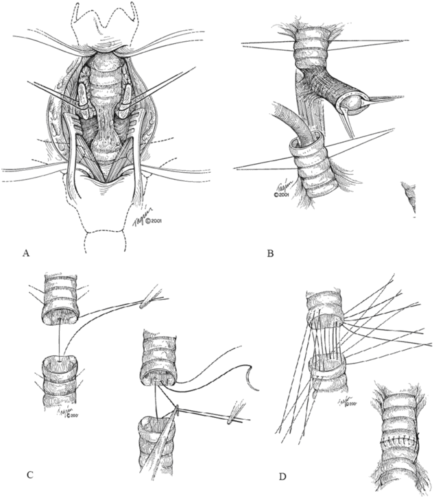
Technique of tracheal resection and reconstruction. The affected portion of the trachea is carefully dissected (A), and cross-field ventilation is used following division of the airway (B). Reconstruction is performed using interrupted absorbable sutures (C), and a tension-free anastomosis is created (D).
Stay updated, free articles. Join our Telegram channel

Full access? Get Clinical Tree


