Severe blunt chest trauma with mediastinal shift to the right (arrows) due to left tension hemopneumothorax.
Focused assessment with sonography for trauma (FAST) is another important adjunct to the primary survey in the patient with a ‘C-problem’. Besides the evaluation for associated abdominal injuries, it is useful to detect pericardial effusion or tamponade in the thoracic trauma patient. Furthermore, thoracic ultrasound not only has the potential to detect intrapericardial bleeding, but in the hands of an experienced investigator it may also allow recognition of hemothoraces and assessment of the heart and its function.
Airways
Consider that airway obstruction may be caused by blood, secretions and foreign objects (e.g. tooth), as well as by injuries of the airway itself or compression from the outside.
Laryngotracheal injury
Despite its protected position between mandible and sternum, the larynx and cervical trachea can be injured by blunt trauma (e.g. hyperextended neck against dashboard in unrestrained passengers, direct blows, strangulation by seatbelt/rope/manually) as well as by penetrating forces (e.g. knife, projectile). Patients may present with stridor, hemoptysis, hoarseness (due to vocal cord injury/displacement or recurrent laryngeal nerve injury, often associated with lesions to the cricotracheal junction) and neck pain. Besides concomitant soft tissue injury (erosion, haematoma), cervicomediastinal emphysema can be present. In stable patients, thorough examination by CT scan and flexible endoscopy allows systematic assessment of the injury, including potentially associated lesions of the esophagus. In patients with severe respiratory distress, oro- or nasotracheal intubation past the area of trauma is advised. Wherever applicable, this is best performed by guidance of a flexible bronchoscope, in order to avoid additional tissue damage with the risk of complete airway disruption. In cases of unsuccessful intubation due to severe edema or concomitant maxillofacial injury, emergency tracheostomy may be required.
Early surgical exploration with definitive reconstruction through a collar incision is recommended. In cases of laryngotracheal lesions with possible bilateral vocal cord and/or recurrent laryngeal nerve injury, consider the placement of a tracheostomy cannula at the end of the procedure.
Tracheal compression by sternoclavicular dislocation
Trauma to the upper chest can result in posterior sternoclavicular joint dislocation or sternoclavicular fracture dislocations, in which the medial end of the clavicle may come to rest on the trachea and cause upper airway obstruction. Amongst inspiratory stridor and a palpable defect in the region of the sternoclavicular joint, patients may also present with signs of vascular compression of the ipsilateral extremity. Treatment consists of immediate reduction of the medial end of the clavicula, best achieved by pulling both shoulders backwards, eventually with the help of a cushion positioned between the shoulder blades, and by additionally pulling at the medial end of the clavicula with a pointed clamp. Once reduction is achieved it is usually stable, and if so, conservative treatment (shoulder immobilization for 3 weeks) is sufficient. In contrast, anterior sternoclavicular dislocations are more common, and reduction is easier, but reduction is rarely stable, and operative fixation is often necessary.
Intrathoracic trachea
Based on the firm constitution of the trachea, especially in its cartilaginous anterior part, rupture is a very uncommon finding. However, if it does happen, it usually occurs due to a sudden increase in intratracheal pressure against the closed glottis, which mainly leads to vertical tears in the posterior, membranous part. These tracheal lesions can also result from forceful intubations. Patients present with haemoptysis, mediastinal emphysema and eventually pneumothorax (uni- or bilaterally), which are generally characterized by large air leaks through an indwelling chest tube. After establishing diagnosis by bronchoscopy and securing the airway by orotracheal intubation, prompt primary repair is advised. Access to the trachea is gained either through a collar incision for cranial lesions with possible extension into an upper partial sternal split or through a right thoracotomy, allowing better exposure of the lower and especially dorsal part of the trachea.
Bronchial rupture
Most bronchial ruptures occur due to shearing forces and forceful antero-posterior compression of the chest, as seen in most car accidents, leading to distracting forces of the laterally displaced lungs at the relatively fixed carina. Most tears occur within 2.5 cm of the carina and are usually found on the right, due to the shorter length and relatively unprotected position of the right mainstem bronchus. Patients may present with only subtle symptoms such as haemoptysis and discrete emphysema if the tear is covered. In tears communicating with the pleural space, pneumothorax with large air leaks through the chest drains may occur and suction on the chest drain may even worse dyspnea. The typical radiological sign is that of a collapsed lung at the bottom of the hemithorax (as opposed to a lung collapse around the hilum seen in ‘conventional’ pneumothorax); therefore it is also referred to as ‘dropped lung’ sign. Again, bronchoscopy establishes the diagnosis, and prompt direct repair through thoracotomy under single-lumen ventilation should be preferred to resectional procedures. Asymptomatic, minor bronchial tears can be managed conservatively in the presence of a fully expanded lung with no air leak being present. In those patients, bronchoscopic examinations should be performed on a regular basis for early recognition of strictures or bronchiectasis, which may develop at a later time in the course of recovery.
Lungs
Pneumothorax
The most common injury resulting from thoracic trauma is a simple pneumothorax. It is either caused by laceration of the visceral pleura by a sharp object (e.g. fractured rib, knife, bullet) or due to rupture of the visceral pleura caused by deceleration or barotrauma. While small pneumothoraces (up to 2 cm) in stable blunt chest trauma victims can be managed conservatively, all patients with pneumothorax and penetrating injury, unstable respiratory situation or otherwise intended intubation and mechanical ventilation need tube thoracostomy. Chest tubes may be removed when the lung is fully expanded (no persistent blood or air in the thoracic cavity), if the air leak has completely resolved and if fluid drainage is less than about 200 ml/24 hours.
The deadliest form of pneumothorax is tension pneumothorax, which develops when a one-way valve-like air leak occurs from the lung. The increasing accumulation of air in the thoracic cavity leads to a complete collapse of the affected lung and a mediastinal shift to the contralateral side with compression of the contralateral lung and venae cavae. The result is a rapidly increasing hemodynamic instability due to decreased venous return, followed by rhythm disturbances (tachyarrhythmia). Patients correspondingly present with chest pain, respiratory distress, tachycardia, hypotension, distended neck veins and a unilateral absence of breath sounds with hyper-resonance to percussion. Sometimes clinical diagnosis can be difficult, since associated hemothorax, lung contusions and hypovolaemia can mask the typical aforementioned clinical signs (Figure 26.1). Radiological confirmation of diagnosis is usually obsolete, and immediate decompression is mandatory by inserting a large-calibre needle through the second intercostal space in the mid-clavicular line. Definitive management consists of insertion of a chest tube in the fifth intercostal space in the anterior axillary line.
Also open pneumothorax can lead to a certain amount of tension in the thoracic cavity if there is a one-way-valve-like tissue flap (or inappropriately placed wound dressing!) that allows air to enter through the opening in the chest with every breath. Management includes coverage of the open chest wound with a three-way occlusive dressing that allows air to go out and at the same time prevents air from entering the thoracic cavity. Then a tube thoracostomy is performed remote from the wound until operative debridement and closure of the defect are possible. Local muscle or myocutaneous flaps should be preferred over prosthetic material (e.g. polypropylene mesh) in the reconstruction process, since wound contamination usually has to be expected.
Hemothorax
Hemothorax can compromise respiration itself on the one hand, and on the other hand, hypotension due to blood loss contributes even more to poor tissue oxygenation. Hemothorax can occur from bleeding due to lung laceration, vessel laceration (usually intercostal or internal mammary vessels) or from fractured ribs, sternum or spine. Small bleeds are self-limiting, and patients with major bleeds, for example, due to hilar vessel injury, heart injury (chamber rupture) or aortic dissection, rarely even reach the hospital alive. Hemothoraces are evacuated using a large-caliber chest tube and the application of autologous blood salvage systems (e.g. Hemovac, Cell Saver) can be helpful in cases of expected large volumes. Operative exploration should be considered if the initial amount of blood output is more than 1,500 ml and/or chest tube output is 200 ml or more for 2–4 hours, also taking into account the patient’s condition. In uncertain situations or if the bleeding source is well localized (e.g. by CT angiography), a videothoracoscopic approach (VATS) can be chosen in hemodynamically stable patients. VATS also is applicable in the post-primary evacuation of large, clotted hemothoraces in order to allow lung re-expansion and prevent the formation of empyema and late fibrothorax.
Lung laceration
Pulmonary lacerations are seen in penetrating as well as blunt thoracic trauma victims and usually result in a combination of bleeding and air leak (i.e. hemopneumothorax). Minor lacerations can be treated by simple insertion of a chest tube. For larger, more centrally located lung injuries (Figure 26.2), characterized by large air leak or persistent blood loss (continuous blood loss of >200 ml per hour), consider thoracotomy. Depending on the degree of injuries, either resectional procedures or, more desirable if applicable, parenchyma-saving techniques, such as pulmonary tractotomy, may be used. This technique initially was developed to avoid larger resectional procedures (i.e. lobectomy, pneumonectomy) in gunshot victims. The basic idea is to open the wound tract by dividing the healthy parenchymal bridge above it between two clamps (or by means of a stapling device), and subsequently bleeding vessels and small, leaking bronchi at the base of the tract can be ligated and oversewn, respectively.
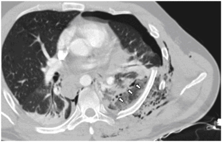
Blunt trauma victim with a large tear across the whole lower lobe (arrows), dislocated rib fractures and pneumothorax.
Lung contusion
Direct lacerations of the lung, as well as the transmitted, indirect forces associated with blunt thoracic trauma, lead to diffuse bleeding and edema in the underlying lung parenchyma (Figure 26.3), resulting in pulmonary shunting (ventilation/perfusion mismatch).
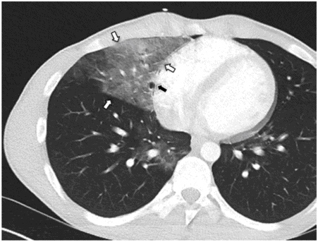
Lung contusion (white arrows) of the middle lobe with small pneumatocele (black arrow).
The degree of respiratory impairment often varies with concomitant injuries (e.g. flail chest, pneumothorax, hemothorax) and pre-existing disabilities (lung emphysema); patients should be closely observed because the full impact on oxygen exchange emerges only hours after the initial trauma. Generally, blood gases deteriorate before radiological signs of lung edema appear; furthermore, these radiological signs might only be minimal if there is a short interval between the triggering injury and radiographical studies, which leads to a high degree of under-estimation in these patients. Therapy includes administration of humidified oxygen, carefully controlled administration of crystalloids (to prevent fluid overload) and close patient observation in order to initiate, if necessary, endotracheal intubation and mechanical ventilation. Haematoma formation can be observed in 5–10% of patients with pulmonary contusions. Symptoms of haemoptysis and occasionally fever usually abate within 1 week, but haematoma resolution on chest radiographs takes about 1 month.
The most severe complication from lung contusion is acute respiratory distress syndrome (ARDS), whereas the risk for ARDS increases with the severity of injuries. Not only the injury to the lung itself but also its combination with associated injuries and the eventual need for endotracheal intubation and ventilation with its risk of ventilator-associated pneumonia finally may result in ARDS.
Chest wall
The thoracic wall protects the vital organs of the chest, notably the heart and lungs, but it also covers the well-perfused and fragile parenchymal abdominal organs of the abdomen such as the liver, spleen and kidneys. Every thoracic injury, whether blunt or penetrating, leaves certain damage to the chest wall. While in children the ribs are more elastic, stronger forces are needed to cause rib fractures, and thus, severe intrathoracic injuries in blunt trauma can occur without signs of broken ribs. Consider that rib fractures in toddlers up to 3 years occasionally result from child abuse. In contrast, in the elderly, simple coughing or a fall from a standing position can result in multiply fractured osteoporotic ribs without severe damage to intrathoracic organs.
Rib fractures
Rib fractures are the main finding in blunt thoracic trauma. Beside pain with the consequence of shallow breathing and atelectasis of the lung, significant respiratory impairment is mostly due to accompanying lung contusions and/or associated head injuries as well as pre-existing co-morbidities (i.e. poor pulmonary reserve). The direct impact on respiratory function as well as the pattern of associated injuries depends on the location (upper/lower ribs, single fracture/fracture in several places) and the number of broken ribs. Since the upper ribs (1,2) are well protected by the clavicles and the whole shoulder girdle, fractures of these ribs result only from strong forces, which should raise suspicion for particular associated injuries such as aortic rupture or tracheobronchial injury.
Fractures of the middle ribs (3–8) are the most common and may result from direct blows to the chest, as well as from indirect lateral fractures following forceful antero-posterior compression of the chest. Furthermore, trauma to the upper extremity often leads to a fracture of the clavicle first, then driving the scapula into the nearby chest, typically causing lateral and posterior fractures of the middle ribs.
Fractures of the lower ribs (9–12), like those of the upper ones, rarely have major influence on respiratory mechanics, but injuries to the liver, spleen and kidneys, as well as diaphragmatic rupture, are sometimes associated with these injuries (Figure 26.4).
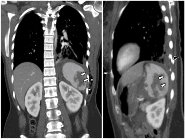
Fracture of lower ribs (black arrow) with associated second-degree splenic laceration (white arrows) and subcutaneus emphysema (stars).
The cornerstone of initial treatment is undoubtedly sufficient pain control to allow appropriate pulmonary toilet. Shallow breathing and the avoidance to cough due to poor pain control inevitably lead to sputum retention, atelectasis and pneumonia. The broad possibilities of pain control reach from oral analgesics, including morphine and its derivatives, over self-administered IV opioids, to epidural analgesia and should be carefully adapted to the patient situation. Operative stabilization of fractured ribs is rarely indicated without the presence of a severe instability of the chest wall due to a series (three or more) of multiply broken ribs, which is called ‘flail chest’.
One of the late sequelae after rib fractures, which may need further treatment, is pseudarthrosis (painful instability at the fracture site 6 or more months after trauma). If local infiltration with long-lasting anaesthetics and corticosteroids does not lead to pain relief and painful instability is one of the main clinical findings, operative measures, such as simple resection or debridement followed by plate osteosynthesis, have to be considered. One of the most difficult and also the most prevalent problems is chronic thoracic pain, which occurs in approximately 30% of patients, and therefore needs an interdisciplinary approach between thoracic surgeons, anaesthetists and on occasion the help of a psychiatrist in the treatment of a post-traumatic stress disorder.
Flail chest
Flail chest is defined as a consecutive series of three or more rib fractures, which are broken in at least two different sites resulting in a free-floating chest wall segment. These patients are in the majority of cases referred after moderate to severe blunt trauma and suffer from respiratory-dependent pain and dyspnoea. In the spontaneously breathing patient, a paradoxical movement of the free-moving chest wall segment can be examined. But often, concomitant haematoma and/or emphysema of the chest wall soft tissue conceal these findings. The flail segment can be anterior, associated with contralateral anterior rib fractures, sternal fractures or separation of several ribs at their costochondral junction (invisible by plain radiographs and even difficult to detect by CT scan). In most cases, the floating segment is localized laterally due to direct impacts, antero-posterior compression or upper extremity trauma (‘scapula against chest wall’ effect). The least common variety lies posterior and often goes clinically unrecognized, since the chest wall in this region is well supported by the back muscles and the scapula.
As flail chest often results in respiratory insufficiency due to a combination of pain, increased work of breathing and, most important, associated pulmonary contusion; endotracheal intubation and ventilation sometimes are inevitable. As internal pneumatic stabilization by mechanical ventilation takes an average ventilation time of 10 days, the risk of ventilator-associated pneumonia as well as the high costs of ICU care are only some of the factors which may raise the question for early rib stabilization. Indications for rib stabilization include:
Thoracotomy for another reason (e.g. haemorrhage or lung laceration)
Severe chest wall instability with increasing respiratory insufficiency in patients with sufficient pain control and without the presence of major lung contusions
A relative indication for chest wall stabilization is an insufficient bronchial toilet due to inefficient coughing in flail chest patients. Roughly summarized, operative stabilization should aim at preventing intubation and prolonged mechanical ventilation. For rib stabilization, contourable reconstruction plates or anatomically precontoured titanium plates are available, which allow anatomical and physiological fracture reconstruction (Figure 26.5). Furthermore, intramedullary splints can be used with smaller incisions to reduce exposure and avoid further soft tissue damage. Implant removal is usually not required.
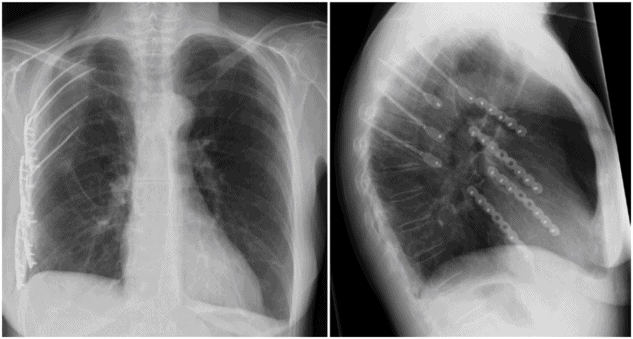
Result after stabilization of flail chest with osteosynthesis plates (lateral fracture line) and splints (latero-posterior fracture line).
Stay updated, free articles. Join our Telegram channel

Full access? Get Clinical Tree


