with Respiratory Failure


Type


Cause


Respiratory drive


 Pharmacological


Drug overdose, anesthesia


 Congenital


Central hypoventilation syndrome


 Acquired


Cerebrovascular accident


Neuromuscular


 Cervical spinal cord injury


Trauma


 Chronic inflammatory demyelinating polyneuropathy


Guillain–Barré syndrome


 Anterior horn disease


Poliomyelitis


 Phrenic nerve injury


Trauma, cardiac surgery


Muscles of respiration


 Pharmacological


Neuromuscular blocks


 Congenital


Hypophosphatemia


 Acquired


Kyphoscoliosis, trauma


Rib cage


 Decreased mobility


Alt. Pleura


 Extrapulmonary restriction


Pneumothorax, pleural effusion


Airway


 Upper airway obstruction


Epiglottitis, foreign body


 Lower airway obstruction


Asthma


Increase in dead space


 Increased ventilation/perfusion ratio


Emphysema


 Decreased ventilation/perfusion ratio


Acute respiratory distress syndrome


 General pulmonary hypoperfusion


Hypovolemia, cardiogenic shock


 Localized hypoperfusion


Thromboembolism


Physiopathology


There are five physiopathological mechanisms that can alter the homeostasis of gases and lead to respiratory insufficiency: (1) an alteration in the alveolar ventilation and pulmonary perfusion ratio (V/Q ratio); (2) a shunt (short circuit); (3) hypoventilation; (4) an alteration in the diffusion of gases in the alveolocapillary membrane; and (5) a decrease in the concentration of inhaled oxygen. The three most important are the V/Q ratio, hypoventilation, and a shunt, with a decrease in inhaled O2 being considered a minor clinical implication of acute respiratory insufficiency.


Ventilation/Perfusion Alterations


An alteration in the V/Q ratio is the most common cause of arterial hypoxemia. The concentration of O2 in the alveoli and capillary blood depends on the relative concentrations of inhaled O2 and unsaturated blood in the pulmonary artery, which is termed the ventilation/perfusion ratio. The same applies to CO2. While it could be expected that there is an adequate ratio for every alveolar unit, the lung does not act as a multitude of identical gas exchange units; rather, it acts as a multitude of parallel and serial perfusion and ventilation units, which vary, even in healthy individuals, because of age, physical activity, bodily posture, and other factors, with some poorly ventilated but well perfused areas, and other areas that are well ventilated but poorly perfused (Fig. 17.1).

../images/441522_1_En_17_Chapter/441522_1_En_17_Fig1_HTML.png

Fig. 17.1

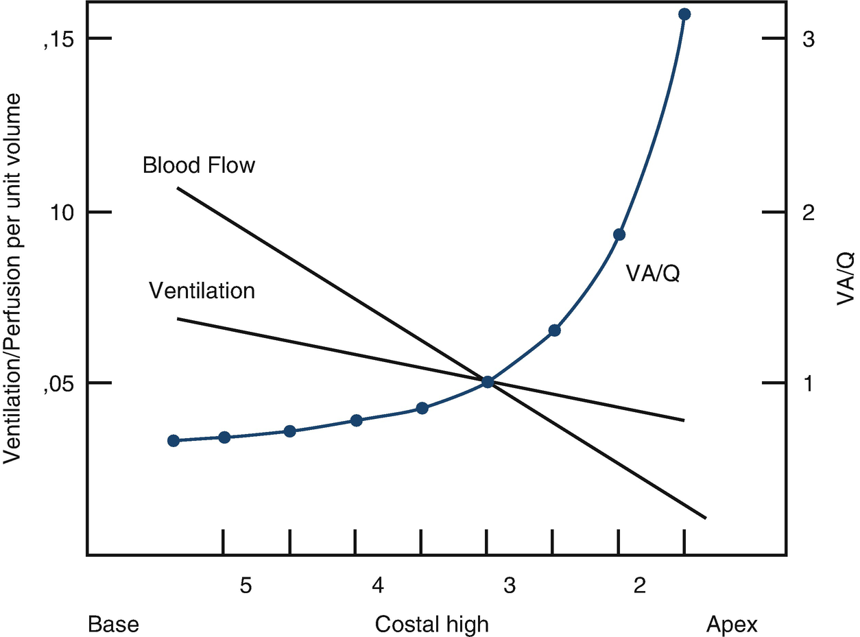
Ventilation distribution, blood flow, and ventilation/perfusion (V/Q) ratio


Alveolar oxygen pressure (PaO2 ) is determined by the pressure of inhaled oxygen, the pressure of alveolar carbon dioxide (PaCO2 ), and the respiratory quotient, while PaCO2 is determined by alveolar ventilation (VA) and the range of corporeal CO2 production. When the pulmonary blood flow of the unit decreases (i.e., the V/Q ratio increases), PaO2 and the capillary oxygen pressure approach the partial inhaled oxygen pressure. When the ventilation of the unit decreases (i.e., the V/Q ratio decreases), PaO2 and PcO2 approximate the PaO2 of mixed venous blood. In the normal lung, the V/Q ratio ranges between 0/6 and 3.0, concentrated mostly at 1.0. Hypoxemia occurs when perfusion exceeds ventilation—that is, when the V/Q ratio is <1 (Fig. 17.2).

../images/441522_1_En_17_Chapter/441522_1_En_17_Fig2_HTML.png

Fig. 17.2

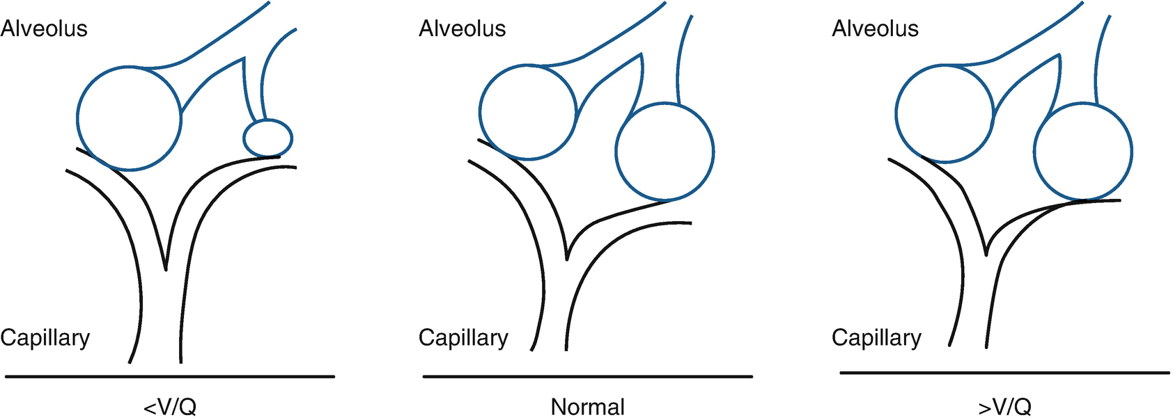
Ventilation/perfusion alteration


In a person who is breathing spontaneously, there are compensatory mechanisms to correct hypoxia and/or hypercarbia. In the context of hypoxemia, pulmonary vascular vasoconstriction consists of the alveolar units with better V/Q ratios an excluding those that are poorly ventilated, while the increase in VA as a response to hypoxia prevents an increase in PcO2, including lowering it to below normal levels.
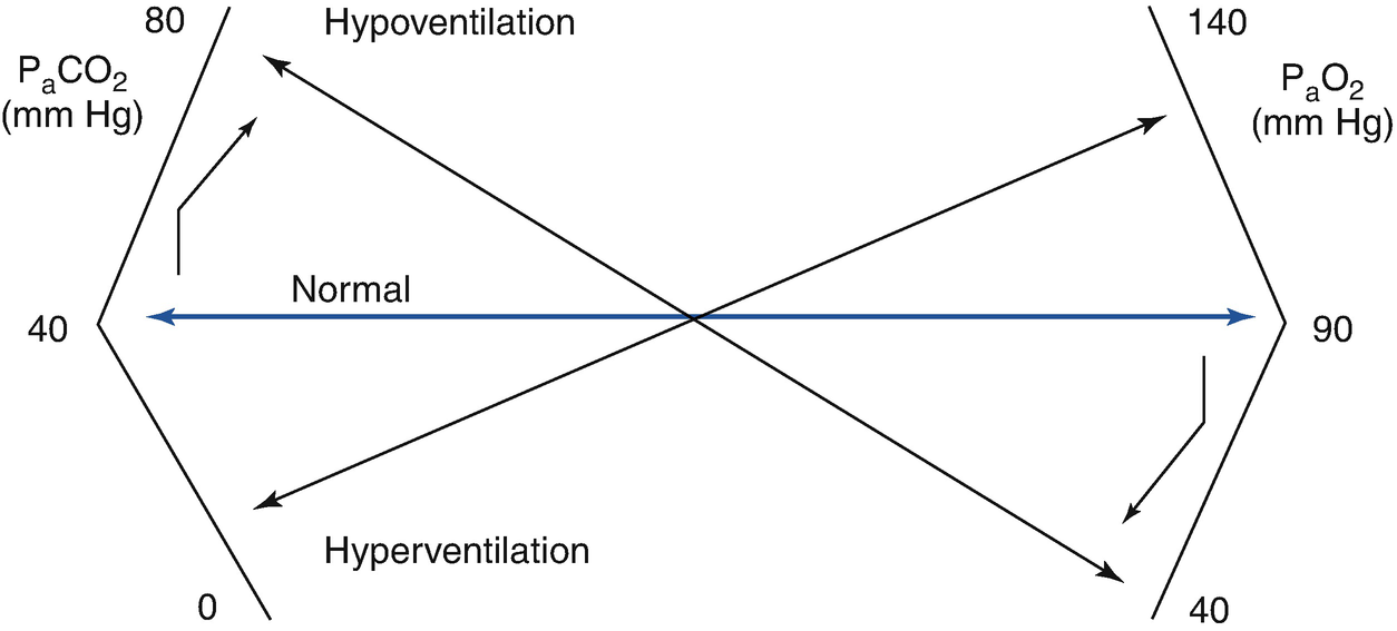

Hypoventilation


PaO2 is determined by the balance between oxygen provided by VA (which provides oxygen from inhaled air) and the extraction of oxygen by capillary blood. Consequently, when VA decreases significantly, PaO2 decreases and PcO2 increases, which are the fundamental characteristics of hypoventilation.


PcO2 is determined by VA and the production of CO2 (VCO2), multiplied by the K constant, as is seen in the formula PaCO2 = (VCO2/VA) × K, from which it can be deduced that increased CO2 production or a decrease in VA increases PaCO2. As the compensatory renal response to hypercarbia is slow (bicarbonate retention), there is an acute fall in arterial pH.


Given that minute ventilation includes both VA and dead space, its reduction or increase implies an increase or decrease in VA. The ratio between the decrease in PaO2 and the increase in PcO2 that occurs in hypoventilation can be calculated by the alveolar gas equation, (PaO2 = PIo2 − (PaCO2/R) + F, where PIo2 is the inhaled oxygen fraction multiplied by barometric pressure, minus water vapor; R is the gas exchange ratio (CO2 production/oxygen consumption), which is determined by the metabolic state of the tissue; and F is a minimal correction. This formula makes it clear that hypoventilation responds to the contribution of oxygen without necessarily decreasing PcO2. The effects of the equation at the arterial level can be seen in Fig. 17.3.

../images/441522_1_En_17_Chapter/441522_1_En_17_Fig3_HTML.png

Only gold members can continue reading. Log In or Register to continue

Stay updated, free articles. Join our Telegram channel

Nov 7, 2020 | Posted by in Uncategorized | Comments Off on with Respiratory Failure

Full access? Get Clinical Tree

Get Clinical Tree app for offline access