Viral Pneumonia

Chapter 25 Viral Pneumonia


Despite increasing recognition of the role of viruses in causing pneumonia in adults, either as sole pathogens or as copathogens with bacteria, diagnosis and treatment of viral pneumonias remain difficult clinical problems. Progress has been made in development of precise laboratory tests to diagnose lower respiratory tract infections of viral origin. Wider availability of such tests is likely to spur recognition and give impetus to development of new therapies.


Burden of Disease


Viral Community-Acquired Pneumonia


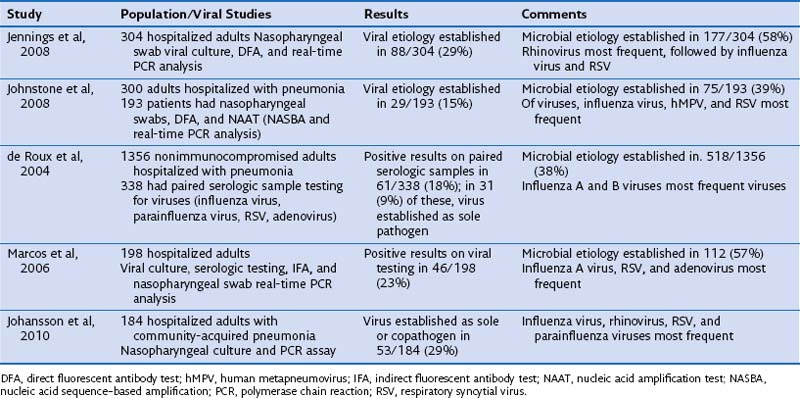
The proportion of pneumonias in adults that are caused by viruses is difficult to quantify with any precision. This lack of certainty is not surprising, because in approximately 50% of pneumonias, an identifiable pathogen cannot be established by conventional laboratory testing. Molecular techniques such as polymerase chain reaction (PCR) analysis improve this number but are rarely performed in routine clinical care. Difficulties in obtaining adequate specimens for viral cultures or molecular testing add to the problem. However, several recent studies have made efforts to quantify the burden of viral community-acquired pneumonias in adults and are summarized in Table 25-1. Although the proportion of viral pneumonias varies in these studies owing to differences in study populations and rigor of testing for viruses, it is clear that viruses cause a significant proportion of pneumonias. Overall, in immunocompetent adults hospitalized with pneumonia, viruses are the responsible pathogens in 15% to 30% of the cases, either by themselves or as copathogens with bacteria. Lower respiratory secretions such as bronchoalveolar lavage (BAL) fluid, tracheal aspirates, or good-quality induced sputum are preferable as specimens for diagnosis of viral pneumonia. Such specimens are more difficult to obtain, however, and most studies have used nasopharyngeal wash samples or swabs for culture and molecular testing. Presence of established pathogens such as influenza virus in such upper respiratory tract samples in a patient with pneumonia most probably implies that the virus is a sole or copathogen in causation of pneumonia; however, this general rule may not be applicable to other viruses. Presence of such viruses as rhinovirus might imply only upper respiratory infection in a patient with pneumonia due to another pathogen. Findings on studies of viral pneumonias must therefore be interpreted with some caution.


Viral Pneumonia in Immunocompromised Patients


Patients who have undergone hematopoietic stem cell transplantation (HSCT) or solid organ transplantation or who are undergoing intensive chemotherapy for leukemias exhibit a higher incidence of viral upper respiratory infections than that in normal control subjects, as well as more frequent involvement of the lower respiratory tract, more severe pneumonias, and higher rates of death from viral pneumonia. All of the pathogens mentioned in Table 25-1 are represented in these patients. Respiratory syncytial virus (RSV), adenovirus, and parainfluenza virus, which usually cause only mild illness in immunocompetent persons, can cause severe pneumonias with high mortality rates in this high-risk patient population. Extrapulmonary manifestations of influenza infection, such as myocarditis, are not unusual in this population. RSV is the most common viral pathogen of the lower respiratory tract in patients who have undergone hematopoietic stem cell or solid organ transplantation, as reported in many series. Fulminant, disseminated adenoviral infections have been reported in patients with advanced human immunodeficiency virus (HIV) disease. Cytomegalovirus (CMV) infection is a major problem in stem cell and solid organ transplant recipients and probably is the most common viral pneumonia among patients who are not on prophylaxis or preemptive treatment regimens.


Pathogens


Although some variability has been documented in the frequency with which different viral pathogens are diagnosed as the etiologic agent of pneumonia in specific series, the most frequent pathogens are influenza A virus, influenza B virus, and RSV. In addition, parainfluenza viruses 1, 2, and 3 and adenovirus, coronaviruses, and rhinovirus also may cause pneumonia. Human metapneumovirus (hMPV) and, more recently, bocavirus have been recognized as pathogens. Rarer pathogens are CMV, varicella-zoster virus (VZV), herpes simplex virus (HSV), rubeola virus (the measles virus), and hantavirus.


Influenza Viruses


The most frequent viral pathogen implicated in causing pneumonia and increased mortality, influenza virus serotypes A and B, especially A, are responsible for outbreaks of respiratory illness each winter. Minor changes in hemagglutinin and neuraminidase proteins of the virus causing “antigenic drift” are responsible for seasonal influenza outbreaks. Although people of all ages are exposed to infection, the consequences of influenza-specific respiratory infection are more severe in certain groups of patients—elderly persons, nursing home residents, infants, those with severe coexisting cardiac and respiratory diseases such as congestive heart failure and chronic obstructive pulmonary disease (COPD), pregnant women, patients with immunosuppression due to hematologic malignancies, and those with HIV disease. Most of the deaths attributable to seasonal influenza occur in these groups. In addition, obesity has been associated with more severe disease and higher mortality in the recent H1N1 pandemic.


Major mutations in hemagglutinin and neuraminidase result in “antigenic shifts” and are responsible for pandemics. The most recently described is the current H1N1 (swine origin) virus pandemic starting in 2009. In contrast with seasonal influenza, the proportion of deaths in younger patients (18 to 65 years) has increased during pandemics. This increased mortality has been attributed to lack of previous exposure to a similar strain of the virus, with corresponding lack of protective antibodies, in patients who were not alive at the time of an earlier outbreak with a similar virus. This pattern has been seen in the current H1N1 pandemic.


Thus, influenza-related pneumonia occurs each year during the fall, winter, and spring seasons as a consequence of infection with circulating seasonal influenza viruses. Distinct from this pattern, pandemic influenza viruses cause pneumonias in explosive outbreaks. Such pandemic viral pneumonias differ from the seasonal disease in several ways: (1) Patients 18 to 65 years of age, especially those in the 20- to 45-year-old age group, are affected more often than in seasonal disease; (2) mortality due to influenza-related pneumonia is increased among adults 20 to 45 years of age; (3) the proportion of primary viral pneumonias is higher than in seasonal disease, in which most influenza-related pneumonias are due to bacteria; (4) viral influenza–related illness occurs outside of the usual influenza season; and (5) special risk factors for severe disease and mortality have been recognized, such as pregnancy and severe obesity. The recent H1N1 pandemic has infected more than 50 million persons in the United States alone, and caused approximately 18,000 deaths worldwide. Severe infections, consisting of pneumonia and acute respiratory distress syndrome (ARDS), typically have been seen in adults, with a median age of 40 years.


The true incidence of pneumonia in influenza respiratory infection is unknown, because most patients with influenza-related illness are evaluated and managed as outpatients without radiographs. Of a group of elderly adults with a high vaccination rate (greater than 90%), 2% to 6% were hospitalized with influenza-related illness; of these, only 5% had radiographically confirmed pneumonia. A significant proportion of such patients will have other illnesses, such as congestive heart failure, that precipitate hospitalization. Pneumonia developing during influenza infection can be one of two types: primary viral pneumonia or postinfluenza bacterial pneumonia. Louria and co-workers, reporting on the 1957-1958 pandemic, described four syndromes of lower respiratory tract illness due to influenza in New York City: (1) influenza illness with physical signs of lower respiratory involvement, but no infiltrates on the chest film; (2) influenza illness followed by bacterial pneumonia; (3) acute, rapidly progressive pneumonia due to influenza virus; and (4) concomitant viral and bacterial pneumonia. The histopathologic changes of influenza-related viral infection of the lower respiratory tract consist of desquamation of tracheobronchial epithelium, alveolar duct dilatation, hyaline membrane covering alveolar surfaces, and hemorrhage and inflammatory infiltrate in the alveolar spaces and interstitium.


Pathogenesis of Bacterial Pneumonia in Influenza


The association of influenza lower respiratory infection with bacterial pneumonia is striking. Such influenza-associated bacterial pneumonias, caused by Streptococcus pneumoniae, Haemophilus influenzae, and Staphylococcus aureus, including methicillin-resistant strains, are more frequent and severe than primary bacterial pneumonias due to the same pathogens. The seasonal incidence of S. pneumoniae infection in a community parallels the incidence of influenza illness. The mechanisms underlying the increased frequency and severity of these influenza-associated bacterial pneumonias are mostly unclear at present, but emerging evidence suggests roles for multiple viral and host factors. Increased apoptosis and desquamation of airway epithelial cells infected with influenza virus exposes multiple surface adhesins for pathogenic bacteria. Influenza viruses expressing PB1-F2, a proapoptotic protein that interferes with mitochondrial function and increases epithelial cell apoptosis, increase susceptibility to S. pneumoniae infection. When infected with viral strains lacking PB1-F2, mice were less susceptible to bacterial pneumonia, compared with those infected with wild-type viral strains. Moreover, influenza infection decreases the efficacy of airway mucociliary clearance, and thus clearance of pathogenic bacteria.


Viral neuraminidase increases bacterial growth and dissemination, and adherence of S. pneumoniae to airway epithelial cells in mouse models. Influenza strains with high neuraminidase activity are associated with higher mortality after bacterial infection. Influenza virus inhibits neutrophil chemotaxis and phagocytic capacity; inhibits, through induction of IFN-γ, macrophage receptor with collagenous structure (MARCO) production by macrophages. MARCO is a scavenger protein involved in macrophage recognition and killing of bacteria. Influenza virus also causes desensitization of Toll-like receptors (TLRs), and this desensitization persists for several weeks.


Jun 12, 2016 | Posted by in RESPIRATORY | Comments Off on Viral Pneumonia

Full access? Get Clinical Tree

Get Clinical Tree app for offline access