Idiopathic Pulmonary Fibrosis and Other Interstitial Lung Diseases

Chapter 47 Idiopathic Pulmonary Fibrosis and Other Interstitial Lung Diseases


Interstitial lung diseases (ILDs) are a large and diverse group of pulmonary disorders that affect the interstitium and air space of the lung and are histopathologically characterized by inflammation and/or fibrosis. Many ILDs are of known cause or association, although the most common remain idiopathic. In 2002 the American Thoracic Society/European Respiratory Society (ATS/ERS) International Multidisciplinary Consensus standardized the classification of the idiopathic interstitial pneumonias (IIPs) and categorized them into seven distinct disorders: (1) idiopathic pulmonary fibrosis (47%-64% of idiopathic ILDs), (2) nonspecific interstitial pneumonia (14%-36%), (3) cryptogenic organizing pneumonia (4%-12%; formerly bronchiolitis obliterans organizing pneumonia), (4) respiratory bronchiolitis interstitial lung disease, (5) desquamative interstitial pneumonia (10%-17%), (6) acute interstitial pneumonia (<2%), and (7) lymphoid interstitial pneumonia (<2%).


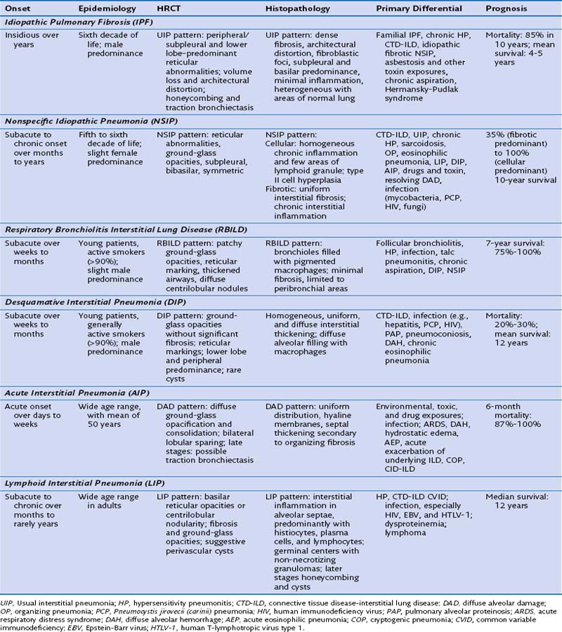
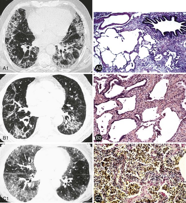
These ILDs differ based on epidemiology, onset of disease, chest imaging findings, histopathology, and most importantly, prognosis and response to treatment. Cryptogenic organizing pneumonia presents and is treated similarly to noncryptogenic presentations of organizing pneumonia, as discussed in Chapter 50. The remaining idiopathic ILDs are discussed in this chapter. Table 47-1 compares various characteristics and Figure 47-1 shows some distinct radiologic and histologic patterns of these idiopathic ILDs.


Idiopathic Pulmonary Fibrosis


Idiopathic pulmonary fibrosis (IPF) is the prototypic chronic, progressive fibrosing interstitial lung disease. In 2011, new ATS/ERS guidelines on the diagnosis, prognosis, and therapy of IPF were published (Raghu et al.). Its prevalence in the United States approaches 100,000 cases with an incidence of 15,000 to 30,000 cases per year. The disease generally affects patients in their sixth decade of life or later, and in patients older than 75 the prevalence increases to 175 per 100,000. There is a slight male predominance.


The cause of IPF is unknown, although risk factors include tobacco use, environmental and occupational exposures, infectious agents such as Epstein-Barr virus (EBV) and hepatitis C virus (HCV), and gastroesophageal reflux disease (GERD). Symptoms are frequently insidious and include cough and shortness of breath, especially with exertion. Symptomatic progression typically occurs over years, although abrupt and otherwise unexplained declines in respiratory symptoms, physiology, and imaging studies (acute exacerbation of IPF) can occur. On examination, digital clubbing may be present. Chest auscultation reveals mid- to end-inspiratory crackles, most pronounced at the bases. During late stages, patients may show evidence of right-sided heart failure caused by the development of pulmonary artery hypertension.


The diagnostic utility of laboratory testing is limited because no biologic fluid-based biomarkers are currently useful in making a definitive diagnosis. Serum Krebs von den Lugen-6 (KL-6), serum surfactant A and D, serum CCL18, serum brain natriuretic peptide (BNP), plasma and bronchoalveolar lavage (BAL) fluid, and matrix metalloproteinases (MMPs) 1 and 7 are all potential biomarkers. Ongoing studies are evaluating the role of these and other potential biomarkers in diagnosis and disease progression. Laboratory testing is useful in defining the clinical context and excluding alternatives to explain the ILD. In particular, serologic testing for connective tissue diseases (CTDs) is recommended in all patients because the distinction between CTD and idiopathic ILD may have prognostic significance. Pulmonary functions tests (PFTs) reveal a restrictive ventilatory defect with a decreased diffusion capacity. BAL findings are nonspecific, and the procedure is not normally required in the diagnostic evaluation. If performed, a mild neutrophilia and mild (<20%) eosinophilia are frequently found. Lymphocytosis, especially greater than 40%, favors an alternate diagnosis.


When a surgical lung biopsy is performed, the histopathologic pattern seen is termed usual interstitial pneumonia (UIP). A definite UIP histopathologic pattern diagnosis requires all four of the following features:


The chest imaging or radiologic pattern seen on high-resolution computed tomography (HRCT) scanning in about 50% of patients is also termed UIP. In the other 50% of patients, less well-characterized patterns are seen. When present, a UIP radiographic pattern has a positive predictive value of 90% to 100% for the presence of a UIP histopathologic pattern. Therefore, when this pattern is present, a surgical lung biopsy is rarely if ever needed. A definite HRCT radiologic pattern diagnosis of UIP can be made if all the following characteristics are present:


In the clinical context of an idiopathic ILD, the identification of a UIP chest imaging pattern confirms the diagnosis of IPF. In patients with discordant findings in their clinicoradiologic appearances, or if they have atypical features on HRCT, a surgical biopsy is recommended in those who need a definitive diagnosis and are suitable for surgery. Transbronchial biopsies are too small to be diagnostic; therefore when a definitive diagnosis is necessary, a surgical biopsy should be obtained, preferably using video-assisted thoracoscopy, with biopsies from more than one lobe. A UIP histopathologic pattern confirms the diagnosis.


In summary, the new guidelines allow the clinician to establish the diagnosis of IPF based on the following criteria:


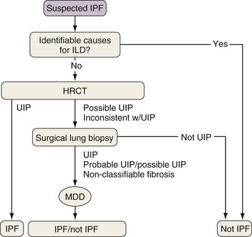
Because the diagnosis of IPF requires combining clinical, radiologic, and often pathologic information, multidisciplinary discussions with chest imagers and pathologists are strongly recommended, especially since a definitive diagnosis cannot be made based on radiologic and histologic findings alone. Figure 47-2 outlines an algorithm for the diagnosis of IPF.


Patients have complications directly and indirectly related to their IPF. Although the natural history of IPF is generally slow symptomatic and physiologic progression, otherwise unexplained rapid deterioration can occur. Acute exacerbations of IPF are defined as the presence of an acute respiratory decompensation of unknown cause and occur in 5% to 10% of patients yearly. Patients tend to present with a viral prodrome and worsening baseline hypoxemia. Invasive mechanical ventilation is often required to maintain oxygenation, but its use is not generally recommended given the poor prognosis associated with acute exacerbations, although it may be appropriate in a minority of patients. Workup should include exclusion of pulmonary embolism, infection, congestive heart failure, volume overload, pneumothorax, and other causes of lung injury. Imaging studies may show new onset of bilateral diffuse ground-glass opacity or dense consolidation. Lung biopsies show a diffuse alveolar damage (DAD) pattern. Although strong evidence is lacking, treatment with high-dose steroids is provided to most patients, in addition to broad-spectrum antibiotics. The prognosis is poor, with mortality rates of up to 70% or more during the following year.


Pulmonary hypertension

Stay updated, free articles. Join our Telegram channel

Jun 12, 2016 | Posted by in RESPIRATORY | Comments Off on Idiopathic Pulmonary Fibrosis and Other Interstitial Lung Diseases

Full access? Get Clinical Tree

Get Clinical Tree app for offline access