and Conor D. Barrett1
(1)
Harvard Medical School Cardiac Arrhythmia Service, Cardiology Division, Department of Medicine, Massachusetts General Hospital, Boston, MA, USA
Abstract
Ventricular tachyarrhythmias have heterogeneous etiologies, clinical consequences, and treatment strategies. Distinguishing ventricular tachyarrhythmias from supraventricular tachycardia (SVT) with aberrancy clinically and on the surface electrocardiogram can be challenging, yet has substantial therapeutic implications. A wide body of randomized clinical trial data has emerged addressing the efficacy of implantable cardioverter defibrillators (ICDs) for the prevention of sudden cardiac death. In this chapter we discuss these issues as well as consensus guideline recommendations for the implantation of ICDs.
Abbreviations
AAD
Antiarrhythmic drugs
ACC
American College of Cardiology
AHA
American Heart Association
CABG
Coronary artery bypass grafting
EPS
Electrophysiology study
HRS
Heart Rhythm Society
ICD
Implantable cardioverter defibrillator
LVEF
Left ventricular ejection fraction
MI
Myocardial infarction
NSVT
Nonsustained ventricular tachycardia
NYHA
New York Heart Association
SVT
Supraventricular tachycardia
VF
Ventricular fibrillation
VT
Ventricular tachycardia
Introduction
Ventricular tachyarrhythmias have heterogeneous etiologies, clinical consequences, and treatment strategies. Distinguishing ventricular tachyarrhythmias from supraventricular tachycardia (SVT) with aberrancy clinically and on the surface electrocardiogram can be challenging, yet has substantial therapeutic implications. A wide body of randomized clinical trial data has emerged addressing the efficacy of implantable cardioverter defibrillators (ICDs) for the prevention of sudden cardiac death. In this chapter we discuss these issues as well as consensus guideline recommendations for the implantation of ICDs.
Distinguishing Wide QRS Complex Tachycardias
A.
History: A prior history of structural or coronary heart disease favors ventricular tachycardia (VT).
B.
Clinical exam: Neither hemodynamic stability nor physical exam findings are of sufficient specificity to be relied upon in order to distinguish between wide QRS complex tachycardia etiologies.
Jugular venous exam may reveal cannon “a” waves due to atrioventricular dissociation in patients with VT.
A third heart sound may favor VT but is not specific enough to diagnose VT.
C.
Differential diagnosis:
Ventricular tachycardia
Supraventricular tachycardia with aberrancy
Preexcitation
Other causes: adverse medication reactions (e.g., digitalis toxicity [specifically associated with bidirectional VT], class Ic agents), ventricular pacing with atrial arrhythmia, metabolic derangement (e.g., hyperkalemia)
D.
The 12-lead ECG may be useful to identify VT from SVT with aberrancy and should be obtained if the patient is hemodynamically stable.
Discrimination of VT from SVT with aberrancy :
Helpful factors are detailed in Table 24-1
Atrioventricular dissociation is characteristic of VT, however given the high prevalence of atrial fibrillation, the absence of evident atrioventricular dissociation does not exclude VT. In some patients with intact VA conduction VTs may have a 1:1 VA relationship, so a 1:1 relationship can not be relied upon to exclude VT and diagnose SVT.
Sensitivity for VT
Specificity for VT
1. Absence of RS complex in all precordial leads (negative precordial concordance)
0.21
1.0
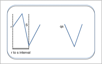
2. R to S interval > 100 ms in any precordial lead
0.66
0.98
3. AV dissociation
0.82
0.98
4. Morphology criteria for VT in V1-2 and V6
0.99
0.97
Overall algorithm
0.97
0.99

Figure 24-1
The rs interval is defined as the duration spanning the onset of the r wave and the trough of the s wave. A qs wave is depicted without any demonstrable r wave
Classification of Ventricular Arrhythmias
Table 24-2
Classification of ventricular arrhythmias
Arrhythmia | Associated features |
|---|---|
Post-infarction ventricular tachycardia (Fig. 24-2) | Mechanism often reentry around scar |
Typically monomorphic unless associated with active ischemia | |
Associated wall motion abnormalities and left ventricular dysfunction on imaging | |
Idiopathic ventricular tachycardia | |
Outflow tract tachycardias (Fig. 24-3) | cyclic adenosine monophosphate mediated delayed afterdepolarizations, triggered activity |
Inferior axis, often left bundle branch block morphology during tachycardia but right bundle branch block morphology may be seen from VT from the left ventricular outflow tract | |
More common in women, can be exacerbated with exercise and in pregnancy | |
Adenosine, calcium channel blocker, or beta-blocker sensitive | |
Treatable with meds or catheter ablation | |
Benign prognosis | |
Fascicular tachycardias (Fig. 24-4) | Reentry involving Purkinje tissue |
Commonest forms are of a right bundle branch block, left anterior fascicular block or left posterior fascicular block pattern during tachycardia | |
More common in men | |
Verapamil sensitive | |
Treatable with verapamil or catheter ablation | |
Benign prognosis | |
Ventricular tachycardia and congenital heart disease | Reentry often involving surgical patch, suture lines, or scar |
May be amenable to ablation but ICDs often favored given structural disease | |
Other ventricular tachycardias | |
Bundle branch reentrant ventricular tachycardia | Reentry involving the bundle branch fascicles |
Typical left or right bundle branch block pattern | |
Associated with structural heart disease (e.g., dilated cardiomyopathy) | |
Poor response to pharmacologic therapy; catheter ablation is first-line therapy; ICDs appropriate as typically unstable | |
Arrhythmogenic right ventricular cardiomyopathy | Reentry around areas of fibrofatty tissue |
Commonly LBBB pattern | |
Baseline ECG | |
May be essentially normal in some | |
1st degree atrioventricular block | |
Epsilon wave (early after depolarization) | |
T wave inversion V1–3 | |
Right bundle branch block or incomplete right bundle branch block, or prolonged S wave upstroke (>55 ms) in V1–3 in absence of right bundle branch block | |
More common in young men, progressive disorder | |
Associated with desmosomal mutations; genetic testing may be useful | |
Typically exercise induced ventricular arrhythmias | |
Sotalol useful, avoidance of competitive sports, ICD in high-risk patients | |
Ventricular tachycardia in nonischemic cardiomyopathy | Reentry around deep myocardial scar or fibrosis |
Polymorphic ventricular tachycardia and fibrillation (Fig. 24-5) | Reentry, automaticity, or triggered activity |
Torsades de Pointes represents “twisting of the points” in context of a long QTc interval | |

Only gold members can continue reading. Log In or Register to continue
Stay updated, free articles. Join our Telegram channel

Full access? Get Clinical Tree


