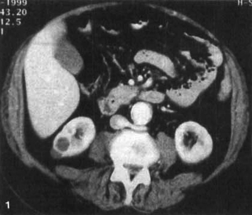
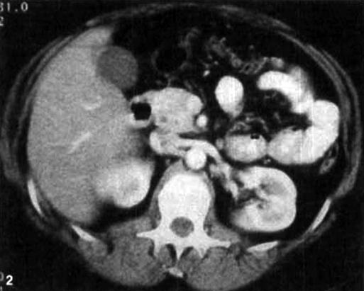
The LRV is an important landmark during open aortic surgery. When it is not found in its normal ventral location anterior to the aorta and posterior to the superior mesenteric artery on preoperative imaging or intraoperative assessment, the surgeon should consider the possibility of a retroaortic venous anomaly. Retroaortic LRV (Figure 1) is seen in approximately 2% of patients and is in itself clinically insignificant. The anomalous renal vein typically courses caudally behind the aorta to enter the IVC at the level of the inferior mesenteric artery. Even when a normal-appearing LRV is seen ventral to the aorta on preoperative imaging or intraoperatively, the presence of structures posterior to the aorta can suggest posterior venous components and possibly a circumaortic LRV or venous collar. Circumaortic LRV or venous collar (Figure 2), the most common renal vein anomaly, is seen in up to 8.7% of patients and carries the most risk for inadvertent injury. The anterior component of the renal collar can easily be mistaken for normal anatomy, whereas the retroaortic component of the renal collar is not readily visible and is easily injured during aortic dissection or clamping. The retroaortic venous components of renal vein anomalies occasionally coalesce with large lumbar and retroperitoneal veins to form a complex retroaortic venous system that is also vulnerable to injury during dissection and clamping.
Venous Anomalies Encountered During Aortic Reconstruction
Retroaortic or Circumaortic Left Renal Vein
![]()
Stay updated, free articles. Join our Telegram channel

Full access? Get Clinical Tree


Thoracic Key
Fastest Thoracic Insight Engine


