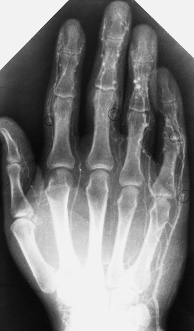
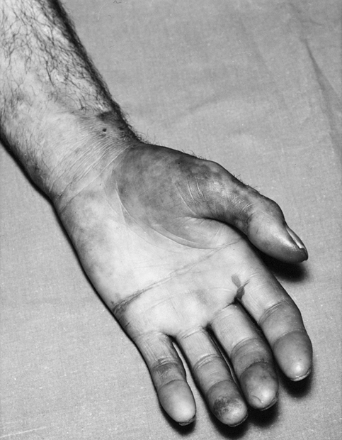
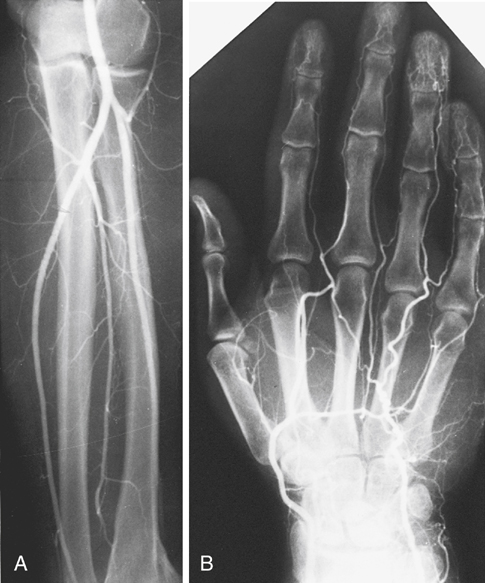
IADI results in injury to the arteries, arterioles, capillaries, and venules. Arterial spasm, embolization of particulate matter, alternations in pH with drug crystallization, platelet aggregation, thromboxane release, and sympathetic-mediated vasospasm have all been proposed to account for ischemic changes. The initial injury appears to affect the venules as the drug and particulate matter pass through the arteriolar and capillary system, then injure venous endothelium, leading to venospasm and venous thrombosis (Figure 1). The venous injury results in outflow obstruction and slowed perfusion, causing stagnation in the arterioles. The result is secondary arteriolar endothelial injury, intimal disruption, and local thrombosis. In addition, outflow obstruction from venous thrombosis increases capillary pressure, leading to increased interstitial pressure and impaired tissue perfusion. The arterial insufficiency, venous injury, and loss of perfusion combine to result in tissue ischemia. Almost without exception, patients present with unrelenting severe pain. Following upper extremity injection, the forearm is often edematous and the hand is usually cool. The fingers are commonly mottled, cyanotic, or gangrenous, with impaired sensation and minimal motor function (Figure 2). Radial and ulnar pulses are usually present, and the presence of intact pulses helps distinguish IADI from other causes of acute ischemia (Figure 3). Although distal pulses are often palpable, plethysmography often fails to identify digital pulses owing to distal arterial occlusion. Initial examination should assess the extent of ischemia and the status of motor and sensory function because the clinical signs and severity of injury correlate with the ultimate outcome and extent of tissue loss. The likelihood of permanent neurologic dysfunction of tissue loss can be more accurately determined through evaluation of the color, capillary refill, temperature, and sensory function of the affected extremity at the time of initial presentation (Table 1). Patients with three or more abnormal findings are significantly more likely to develop neurologic dysfunction or tissue loss than those with one or two abnormal signs if treatment is not initiated within 48 hours of injury. TABLE 1 Clinical Findings Evaluated in Assessing Extent of Ischemia after Intraarterial Drug Injection From Treiman GS, Yellin AE, Weaver FA, et al: An effective treatment protocol for intraarterial drug injection, J Vasc Surg 12:456–465, 1990. The largest experience with a defined treatment protocol comes from Los Angeles County–University of Southern California Medical Center, with its effectiveness on 69 patients reported in 1990. In that study, patients were treated with IV heparin sodium 10,000 units, followed by a continuous heparin infusion adjusted to keep the partial thromboplastin time (PTT) at 2 to 2.5 times control, dexamethasone 4 mg IV every 6 hours, and low-molecular-weight dextran (dextran-40) by IV infusion at 20 mL/hour (Box 1). Narcotics were given as required for pain control, the extremity was elevated in an Osborne sling, and early active and passive range of motion were instituted. The protocol was continued for a minimum of 48 hours and continued thereafter until symptoms completely resolved or extremity function no longer improved. Débridement of nonviable or gangrenous tissue was delayed until there was demarcation to allow preservation of as much potentially viable tissue as possible. Patients managed with this protocol with one or two abnormal findings had an excellent prognosis, regardless of the time from injury to the initiation of treatment (Table 2). For patients with more than two abnormal findings, time to initiation of treatment was important. This protocol was not based on randomized clinical data and has not been compared with other management schemas. Nevertheless, early initiation was found to be effective in preventing tissue loss and permanent neurologic dysfunction in patients with more than two abnormal signs. TABLE 2 Effect of Clinical Findings and Time to Treatment on Outcome at Los Angeles County–University of Southern California Medical Center
Vascular Injury Secondary to Drug Abuse
Arterial Injury
Pathophysiology

Presentation


Clinical Finding
Normal
Abnormal
Color
Pink
Cyanotic
Capillary refill
<3 sec
>3 sec
Temperature
Warm
Cool
Sensory deficit
Present
Absent
Treatment
Number of Abnormal Findings
Time to Treatment (hours)
Normal
Abnormal
p Value
One or two (24 patients)
≤24
17
1
<.001
>24
5
1
<.001
Two or more (24 patients)
≤24
10
2
<1.00
>24
0
12
<1.00 ![]()
Stay updated, free articles. Join our Telegram channel

Full access? Get Clinical Tree


Vascular Injury Secondary to Drug Abuse
