Despite their potential as a sensitive measure of ventricular performance, tissue Doppler velocities vary with normal aging. This is inconvenient for nonspecialists to interpret and makes it difficult to use as an entry criterion for clinical studies. The age-adjusted tissue Doppler Z-scores might avoid these disadvantages and be more discriminant for myocardial impairment than the raw velocities. We conducted a meta-regression of studies reporting age-specific normal tissue Doppler velocities to determine a consensus formula for Z-scores (8 studies, 1,867 patients) that we then tested in an independent study at our institution. We next compared the Z-scores head-to-head with the raw velocities for their ability to distinguish a fresh set of 81 healthy subjects from groups in whom subtle ventricular dysfunction might be expected, including 50 patients with dilated cardiomyopathy, 50 with aortic regurgitation, and 50 with mitral regurgitation. The discriminant capacity, assessed by the area under the receiver operating characteristic curves, was higher for the Z-scores than for the raw velocities in each patient group. At the septal angle of the mitral annulus: dilated cardiomyopathy 0.95 versus 0.92 (p = 0.03), aortic regurgitation 0.83 versus 0.78 (p = 0.02), mitral regurgitation 0.85 versus 0.81 (p = 0.04). At the lateral angle: dilated cardiomyopathy 0.94 versus 0.88 (p = 0.005), aortic regurgitation 0.92 versus 0.83 (p = 0.001), mitral regurgitation 0.87 versus 0.85 (p = 0.31). In conclusion, the Z-scores of the tissue Doppler velocities were better than the raw velocities at detecting myocardial impairment in valvular or heart muscle disease. The calculation needs only the raw velocity and patient age. Tissue Doppler Z-scores could be used to create a novel, more sensitive, definition of ventricular dysfunction and might make it easier for nonspecialists to interpret the reports.
Diagnosing diastolic dysfunction can be challenging. The theoretical reference standard is invasive measurement of the pressure-volume loops, but this is impractical for routine application. Of the noninvasive measures, tissue Doppler imaging (TDI) has been demonstrated to be a reproducible, sensitive, and specific indicator of diastolic dysfunction. It is relatively preload independent compared to the transmitral flow indexes and correlates better with invasive indexes than other echocardiographic measures of diastolic function. The interpretation of tissue Doppler velocities requires an extra step (not needed for the ejection fraction) because the early diastolic (E′) velocity, the main measure of diastolic function, decreases with age. Currently, no convention has been determined for how to account for this. Although a useful rule of thumb is that the normal values for E′ are >10 cm/s, it is clearly not ideal to apply this in a blanket fashion, especially for younger subjects. This age variability is particularly difficult for nonimaging specialists and noncardiologists to account for. Our study examined the feasibility and potential clinical utility of calculating age-specific Z-scores rather than the raw velocities for tissue Doppler indexes of diastolic function. In addition to the raw E′ velocity, such a Z-score would describe how many standard deviations the velocity differed from the age-standardized norm.
Methods
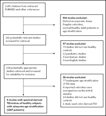
Two investigators (H.Y. and K.M.) performed the literature search and reviewed all the results. PubMed (National Library of Medicine) was used as the bibliographic database. All studies analyzing healthy patients for tissue Doppler indexes of cardiac function with subdivision by age were included. The search terms were (“Diastole” OR “diastol*” OR “diastole” “diastolic”) AND (“tissue doppler” OR “TDI” OR “tissue-doppler” OR “tissue doppler*” OR “doppler tissue”) AND (“Age Factors” OR “Age Distribution” OR “Age Groups” “Age of Onset” OR “Age” OR “age*” OR “aging”). Only English-language studies since 1990 were included. The reference lists of review articles, where appropriate, were searched to identify other potentially eligible studies.
Full articles were retrieved if the abstracts suggested the following criteria had been met: (1) healthy volunteers were studied; (2) an age-based breakdown of data was provided; and (3) spectral-derived tissue Doppler indexes were recorded. Pediatric studies were excluded.
The data were then extracted from the relevant studies. This included demographic breakdowns, 2-dimensional echocardiographic data, and tissue Doppler indexes. Variance-weighted regression analysis was used to calculate the average slope and intercept for the change in E′ with age. Individual linear regression analysis was performed for the change in the E′ standard deviation with age, and a weighted average of the slope and intercept, according to study size, was calculated. In total, 8 studies met our selection criteria, incorporating 1,867 patients in the age range of 20 to 80 years. A flow chart of selection criteria is presented in Figure 1 .
We studied 81 volunteers, aged 15 to 88 years, as controls. No patients had any symptoms suggestive of cardiac disease, none had modifiable risk factors for coronary artery disease (hypertension, diabetes, hypercholesterolemia), and none were taking medication. All had normal electrocardiographic and 2-dimensional echocardiographic findings. We then searched the clinical and echocardiographic records of outpatients at St. Mary’s Hospital, London from 2005 to 2008. A total of 50 patients with moderate, moderate-severe, or severe aortic regurgitation with associated systolic dysfunction, 50 patients with moderate, moderate-severe, or severe mitral regurgitation with associated systolic dysfunction, and 50 patients with dilated cardiomyopathy were included in the present study. The assessment of the severity of valvular heart disease was based on the combined analysis of global left ventricular function and structure and Doppler studies. All patients with dilated cardiomyopathy had either normal coronary angiographic findings or normal stress echocardiographic findings.
Each study participant underwent echocardiography using conventional 2-dimensional, pulsed wave, and TDI techniques. The patients were placed in the left lateral decubitus position and scanned using a Sonos 7500 and IE33 Philips Medical Systems (Andover, Massachusetts) echocardiographic ultrasound imaging system. Both parasternal and apical imaging windows were achieved using a S5-1, 3.5-MHz transducer at a mean depth of 16 cm. Myocardial tissue Doppler peak systolic (S′), early diastolic (E′), and late diastolic (A′) velocities were measured (in cm/s) with the sample volume positioned at the septal and lateral angles of the mitral annular ring. The pulsed wave TDI sample volume length for each patient was set at 2 to 5 mm to minimize spectral broadening. All TDI measurements were taken by a single investigator (H.Y.) and averaged for 4 consecutive beats.
All values are expressed as the mean ± SD or as percentages. All continually variable data underwent tests for normality using the Shapiro-Wilk test and the Kolmogorov-Smirnov test with Lilliefors significance correction. Comparisons between groups were made using Student’s t test. A comparison of the area under the receiver operating characteristic (AUROC) curves were performed using the method detailed by DeLong et al. Linear least squares regression analysis was performed to assess the relations between variables. p Values <0.05 was considered statistically significant. Excel (Microsoft, San Jose, California) and MedCalc (Frank Schoonjans, Mariakerke, Belgium) were used for statistical analysis.
Results
Eight studies, incorporating 1,867 patients, published from 1997 to 2008 were included ( Table 1 ). The data in each study underwent linear regression analysis for the change in E′ and the standard deviation of E′ with age. For the final regression equation, a weighted mean of the gradient and intercept was then produced for E′ (variance weighted) and the standard deviation of E′ (study size weighted). The Z-score of E′ velocity was calculated as the difference between the patients’ E′ velocity and the age-modeled E′ velocity, divided by the standard deviation for that age (i.e., how many standard deviations from the norm was the patient’s E′ velocity). The regression equations and a worked example are shown in Figure 2 .
| Study | Subjects | Septal | Lateral | ||||||
|---|---|---|---|---|---|---|---|---|---|
| Mean of E′ | SD of E′ | Mean of E′ | SD of E′ | ||||||
| Gradient | Intercept | Gradient | Intercept | Gradient | Intercept | Gradient | Intercept | ||
| Munagala et al, 2003 | 1,012 | −0.07 | 12.9 | −0.02 | 3.5 | −0.15 | 19.8 | −0.04 | 5.7 |
| Wierzbowska-Drabik et al, 2008 | 80 | −0.15 | 19.3 | −0.05 | 4.7 | −0.16 | 21.5 | −0.06 | 6.4 |
| Alam et al, 1999 | 62 | −0.14 | 19.5 | −0.02 | 3.2 | −0.20 | 25.6 | 0.02 | 1.5 |
| Sohn et al, 1997 | 59 | −0.13 | 15.9 | 0.00 | 1.5 | ||||
| Van de Veire et al, 2006 | 249 | −0.09 | 13.5 | 0.00 | 2.3 | ||||
| De Sutter et al, 2005 | 174 | −0.10 | 13.8 | −0.02 | 3.0 | ||||
| Bukachi et al, 2008 | 128 | −0.20 | 25.6 | −0.04 | 5.7 | ||||
| Tighe et al, 2003 | 103 | −0.19 | 24.7 | −0.01 | 4.0 | ||||
| Summary | 1,867 | −0.105 | 14.5 | −0.017 | 2.89 | −0.17 | 21.9 | −0.036 | 5.28 |
The summary patient demographic and echocardiographic data are listed in Table 2 . The 81 healthy controls, 50 patients with aortic regurgitation (24 with moderate, 11 with moderate-severe, and 15 were severe), 50 patients with mitral regurgitation (13 with moderate, 25 with moderate-severe, and 15 with severe), and 50 patients with dilated cardiomyopathy were recruited.
| Characteristic | Controls (n = 81) | Dilated Cardiomyopathy (n = 50) | Mitral Regurgitation (n = 50) | Aortic Regurgitation (n = 50) |
|---|---|---|---|---|
| Age (years) | 51 ± 28 | 55 ± 12 | 51 ± 13 | 55 ± 14 |
| Men (%) | 66% | 72% | 64% | 72% |
| Left ventricular end-diastolic dimension (cm) | 4.6 ± 0.7 | 6.0 ± 1.3 | 5.5 ± 0.8 | 5.3 ± 0.8 |
| Left ventricular end-systolic dimension (cm) | 3.2 ± 0.4 | 5.2 ± 1.3 | 4.3 ± 1.0 | 3.8 ± 1.0 |
| Intraventricular septum in diastole (cm) | 1.1 ± 0.3 | 1.1 ± 0.3 | 1.1 ± 0.3 | 1.3 ± 0.3 |
| Intraventricular septum in systole (cm) | 1.4 ± 0.2 | 1.3 ± 0.3 | 1.3 ± 0.3 | 1.6 ± 0.4 |
| Fractional shortening (%) | 33 ± 4 | 15 ± 10 | 25 ± 11 | 29 ± 11 |
| Ejection fraction (%) | 58 ± 6 | 30 ± 14 | 40 ± 20 | 51 ± 15 |
| Left atrial diameter (cm) | 3.6 ± 0.4 | 4.5 ± 0.6 | 4.7 ± 0.6 | 4.2 ± 0.9 |
| Deceleration time (ms) | 202 ± 33 | 189 ± 80 | 202 ± 84 | 223 ± 58 |
| Mitral E (cm/s) | 75 ± 24 | 79 ± 32 | 109 ± 41 | 90 ± 44 |
| Mitral A (cm/s) | 66 ± 22 | 65 ± 26 | 85 ± 33 | 85 ± 38 |
| Mitral E/A ratio | 1.27 ± 0.57 | 1.27 ± 0.66 | 1.38 ± 0.64 | 1.17 ± 0.61 |
Stay updated, free articles. Join our Telegram channel

Full access? Get Clinical Tree


