Urologic Surgery

Chapter 73 Urologic Surgery


Urology as a specialty discipline has much in common with general surgery. Our focus on abdominal and retroperitoneal anatomy, surgical approach, combined use of endoscopic, laparoscopic, robotic, and open surgical technologies, and remarkable diversity of disease processes connects us in the surgical sciences. Urologists work closely with general surgeons regularly, and can profit greatly from each other’s skills and knowledge.


During my experience as Chief of Urology at Ben Taub General Hospital in Houston over the past 20 years, I have had the great privilege of enjoying an outstanding collaborative relationship with the superb general, vascular, and trauma surgeons at our institution. We have worked closely under the stress of major trauma surgery, complexity of exenterative surgery for advanced pelvic malignancies, management of iatrogenic urologic and surgical injury, and challenges of necrotizing infections of the genitalia and perineum.


The surgical resident in training, while moving along the path to becoming a competent general surgeon, must acquire a thorough understanding of urologic disease and urologic surgery. This is equally important for those who practice in settings in which a urologist is likely unavailable, as well as in the setting in which appropriate consultation, surgical planning, and regular operative collaboration are routine.


The following discussion is intended to provide a fundamental overview of urologic surgery for the general surgery resident. As your colleagues in the house of surgery, I welcome and encourage your in-depth pursuit of the fascinating history and practice of urology as your career unfolds.


Urologic Anatomy For The General Surgeon


It is essential for the general abdominal surgeon to be intimately familiar with genitourinary anatomy to complete standard general surgical procedures, avoid iatrogenic injury, and deal with abnormal anatomy.1 Examples of particular areas of risk or challenge include the renal vasculature in major vascular and retroperitoneal tumor surgery, ureteral mobilization and injury avoidance in complex colonic procedures, urethral and prostatic relationship to the rectum, and spermatic cord and scrotal contents in complex inguinal hernia surgery.


Upper Abdomen and Retroperitoneum


The kidneys are located anterior to the psoas and quadratus lumborum muscles; adjacent to the left kidney are the body and tail of the pancreas, spleen, splenic flexure of the colon, and colonic mesentery. On the right, the kidney is adjacent to the liver, duodenum, and head of the pancreas. In the setting of neoplastic or inflammatory processes involving any of these structures, or the kidney, complex adhesions can form that create challenging dissection planes and increase the risk of organ injury with surgical exploration or mobilization. Although the perirenal fascia (Gerota’s fascia) separates the kidney capsule and parenchyma from these adjacent organs and reduces the risk of renal injury with local dissection, renal parenchymal injury is possible with abnormal anatomy. In the setting of inflammatory disease, the Gerota’s fascia may be densely adherent and inseparable from the kidney, requiring an additional approach to renal mobilization to avoid capsular or parenchymal injury. In the setting of retroperitoneal trauma, it is easy to strip the renal capsule from the parenchyma inadvertently when the kidney is rapidly dissected digitally; this maneuver is therefore best done with care and under direct vision whenever possible, because preserving the capsule enhances renal reconstructive efforts.


The renal pedicle has a predictable anatomic orientation, although anomalies of the venous system and the renal collecting system are not uncommon and can confuse the operating surgeon. The renal vein is typically most anterior, the renal artery just posterior to the vein, and the renal pelvis and ureter most posterior. On the left side, the renal vein typically passes anterior to the aorta and inferior to the superior mesenteric vein as it courses toward the vena cava. On the right, the renal vein can be short and enters the vena cava laterally. The major branches of the renal vein are the gonadal vein on the left (the right gonadal vein enters the vena cava directly), the adrenal vein, which joins the left renal vein superiorly on the left, and enters the vena cava directly and at times posteriorly on the right, and variable lumbar veins, which may be found entering the renal veins posteriorly, coursing directly posterior just lateral to the aorta on the left, or near the vena cava on the right. These major branches are particularly prone to injury with dissection of the renal pedicle and can result in troublesome bleeding. It is the presence of these predictable renal vein branches that allows ligation of the left renal vein near the vena cava in the trauma setting, with preservation of renal venous drainage in most cases. On the right, ligation of the renal vein will typically result in renal vein thrombosis and renal infarction. Renal vascular anomalies for which the surgeon must beware include the circumaortic or retroaortic renal vein on the left and multiple renal veins or arteries on either side. Congenital anomalies involving the kidneys and upper urinary tract may result in unintended injury if not appreciated preoperatively. Renal malrotation, duplication, and fusion anomalies are of particular concern. A pelvic kidney could be removed if thought to represent a pelvic mass of neoplastic origin and the isthmus of a horseshoe kidney might be injured if not appreciated prior to exposure to the great vessels in retroperitoneal surgery, because it invariably crosses anterior to the aorta, just inferior to the inferior mesenteric artery. Renal ectopia is accompanied by markedly variable and unpredictable renal vasculature, with multiple branches, possibly arising from the iliac arteries or aortic bifurcation. Duplication of the renal collecting system with a supernumerary renal pelvis and ureter is notorious for precipitating iatrogenic injury; the unsuspecting surgeon might be caught unaware after identifying a single ureter on either side.


Ureter


The ureter travels from the renal pedicle to the bladder hiatus in a predictable manner, but can be difficult to identify when the region is anatomically hostile because of prior surgery, hematoma, fibrosis, inflammation, or neoplasm. The ureter is found lateral to the aorta or vena cava in the upper abdomen, typically just anterior to the psoas muscle. It then courses laterally, passing over the iliac vessels at the level of the bifurcation of the common iliac into the external and internal iliac branches. The ureter then follows the lateral pelvis, extending deep into the pelvis and passing posterior to the vas deferens in the male and behind the uterine vessels in the female pelvis, before penetrating the detrusor muscle of the bladder posterolaterally. The ureter is best identified in an area of normal anatomy and then followed to the area of concern. This is readily accomplished medial to the lower pole of the kidney or at the iliac bifurcation. When mobilizing the ureter, it is important to avoid undue traction, which could tear or avulse the ureter when it is friable or fixed by surrounding fibrosis, and to achieve the proper dissection plane to avoid devascularization, which is outside the adventitial sheath. The primary longitudinal blood supply to the ureter is found between the muscularis and the adventitia; it is important to preserve this vascular network. The blood supply to the upper third of the ureter arises mainly from branches of the renal artery and small direct branches of the aorta. The middle third of the ureter draws its blood supply from small iliac branches and, at times, branches from the gonadal vessels. The lower third of the ureter has blood supply from gonadal, hemorrhoidal, internal iliac, and superior and inferior vesical branches. In the female, uterine and cervical vessel branches support the ureter, as do vasal branches in the male. Following prior surgery or retroperitoneal disease processes, any of these rich collateral blood supply sources may not be contributory; thus, it is critical to avoid unnecessary extensive circumferential dissection of the ureter and be cognizant of the potential to cause ischemic injury with injudicious dissection, particularly if the longitudinal vessels are disrupted and the branches from the deep pelvis are unreliable following prior insults to the ureteral feeding vessels.


Pelvic Anatomy: Bladder, Prostate, and Seminal Vesicles


The bladder has a rich adventitial layer covered by the pelvic peritoneum along the dome and upper posterior wall, under which lies the detrusor muscle, with the submucosa and mucosa internally. The blood supply to the bladder comes from the superior and inferior vesical arteries, which are branches of the internal iliac artery. The prostate lies just caudal to the bladder neck and gains blood supply from the inferior vesical artery branches. Anterior to the lower bladder segment and the prostate lies the dorsal venous plexus (Santorini’s plexus), which runs behind the pubic symphysis and is contiguous with the dorsal venous system of the penis or the clitoris in the female. The anterior and lateral bladder walls do not have a peritoneal surface but reside within pelvic fat or may abut the musculature of the pelvic sidewall and pubis anteriorly. The bladder can be injured when entering the abdomen through a midline incision if care is not taken to enter the superior retropubic space (of Retzius) and displace the bladder posteriorly with simple blunt dissection when extending the midline rectus fascial incision to the pubis. By transperitoneal palpation, the balloon of a Foley catheter can be palpated within the bladder, ensuring its correct position and confirming that the bladder is well-drained.


When performing complex pelvic surgery in which the bladder is adherent to a pathologic process, such as diverticulitis or sigmoid colon or rectal cancer, I recommend having full sterile access to the urinary tract, with the genitalia prepped into the operative field and any urinary catheters inserted in a sterile fashion. This allows easy intraoperative filling and drainage of the bladder to aid in its identification and also supports the efforts of a consulting urologist if the bladder needs to be entered, repaired, or partially excised. This may be followed by changing to a larger bore catheter to encourage drainage postoperatively.


When ureteral catheters are placed prior to abdominal surgery to aid in ureteral identification, these catheters are typically tied to the exiting Foley catheter with sutures, allowing all catheters to drain during the operative procedure and enabling ready manipulation of all the tubes intraoperatively. In pelvic trauma, particularly from penetrating injuries, when gross hematuria is present, it is sometimes most expeditious to open the bladder via a longitudinal anterior cystotomy incision to perform inspection and repair. The efflux from the ureteral orifices can be observed through this approach, the entire surface can be evaluated, and the entire bladder is accessible through this exposure, which is easily closed, with minimal morbidity. The prostate and seminal vesicles are typically encountered by the general surgeon during pelvic exploration for trauma or during abdominoperineal resection for rectal cancer. Following pelvic radiotherapy and/or chemotherapy, the dissection plane between the Denonvilliers’ fascia, which is just posterior to the prostate, may become fibrotic or obliterated, causing the surgeon to be at risk to enter the prostatic parenchyma or bulbomembranous urethra while trying to separate the perirectal tissues from the urinary tract. When recognized early, the correct dissection plane can be reestablished, although prostatic and urethral repair may be challenging if tissues from the genitourinary side of this dissection are excised or extensively damaged before recognition of the errant plane of dissection. The presence of the Foley catheter is most helpful in guiding the proper dissection, but does not eliminate the potential for iatrogenic injury. The seminal vesicles extend superolaterally from the base (cephalad extent) of the prostate and are prone to injury in the same situations as noted; if entered, they can be sutured or excised with little concern, unless fertility is an issue for the patient.


Groin, Genitalia, and Perineum


Inguinal anatomy is well known to all general surgeons. The relevant urologic issues in this anatomic region relate to the spermatic cord and male genitalia. The detailed anatomy of the scrotal contents is beyond the scope of this discussion; suffice it to say that the testis lies within a mesothelial envelope, the tunica vaginalis. The vascular and genital ductal structures leave the testis from the mediastinum in the posterosuperior portion and travel via the scrotal neck into the inguinal canal. The spermatic cord is invested by the internal spermatic fascia, derived from the transversalis fascia. The cremaster muscle is derived from the internal oblique. The external spermatic fascia, derived from the external oblique aponeurosis, joins the cord below the external ring. The cord is susceptible to injury during inguinal dissection for hernia repair, especially in redo cases, when it may be encased in fibrosis and injured without recognition. Significant injury to the cord may put the viability of the testis at risk, even though the testis is supported by three collateral blood supply sources—external spermatic artery from the external iliac, internal spermatic or gonadal artery from the aorta, and vasal artery from the internal iliac branches. The vas deferens itself may also be injured anywhere along its course, from the midscrotal level to the inguinal canal to its intrapelvic portion. When inguinal herniorrhaphy is performed, care must be taken to avoid excessive tightening at the internal ring level, because compression of the cord at this level can result in venous, lymphatic, or arterial obstruction. In addition, the common use of nonabsorbable mesh materials in hernia repair, whether performed through an open inguinal or laparoscopic approach, may result in fibrotic and inflammatory changes involving the vas deferens, which can cause late occlusion and potentially azoospermia and infertility. This phenomenon has been well-documented in the literature; patients in their reproductive years should be counseled about this risk of groin surgery.2


At times, exploration for inguinal hernia may reveal other forms of scrotal pathology that were misdiagnosed preoperatively, including hydrocele and spermatocele. Urologic consultation may be helpful when atypical groin or scrotal anatomy is anticipated preoperatively or encountered at the time of surgery.


The evaluation of the acute scrotum requires a sophisticated knowledge of genital structure and normal anatomy, as well as extensive clinical experience, for accurate diagnosis. The epididymis is located posteriorly and slightly lateral position to the testis; distinguishing acute epididymitis or epididymo-orchitis from testicular torsion or trauma is a commonly encountered clinical challenge. Scrotal ultrasound may be a helpful adjunct to history and physical examination in the rapid assessment of patients,3 because it is critical to rule out testicular torsion promptly to avoid permanent ischemic injury.


Penile anatomy is seldom relevant to the general surgeon, aside from issues related to urethral catheterization. Urologists are well versed in penile and urethral surgery in the elective and emergency settings and can be helpful to the general surgeon when urethral catheterization is challenging, in the trauma setting, and in the setting of neoplastic and inflammatory disease when anatomy is obscure.


In perineal surgical approaches, urology consultation may be helpful to the general surgeon, as noted, when performing abdominoperineal resection for rectal cancer and when performing incision and drainage or débridement for a perirectal abscess, perineal gangrene, or local neoplasms. The male and female urethras are palpable throughout their length, from the tip of the penis to the prostate in the male or from the urethral meatus to the bladder neck in the female. In the male, the bulbomembranous and prostatic urethra are immediately adjacent to the sites of dissection for these entities. The presence of a urethral catheter is helpful in palpating the location of the urethra, but the corpus spongiosum surrounding the bulbar urethra and the prostatic urethra and parenchyma are still vulnerable to injury with dissection in an errant or obliterated anatomic plane. At the level of the membranous urethra, within the external sphincter of the pelvic floor, the corpus spongiosum essentially disappears, leaving only the relatively thin-walled urethra, which must be approached with great care to avoid inadvertent laceration, transection, or excision.


Endoscopic Urologic Surgery


Urology was the first surgical specialty to embrace diagnostic and therapeutic endoscopic surgery. The history of the development of the cystoscope and the modern evolution of ureteroscopy, percutaneous nephroscopy, and urologic laparoscopy represent fascinating stories in the book of surgical history. Urology as a discipline has the good fortune of being able, in most situations, to assess the urinary tract fully with endourologic techniques and high-resolution imaging; thus, in urology, exploration of the various components of this organ system, without a reasonably well-formulated preoperative diagnosis, is uncommon. The general surgeon should have a basic working knowledge of urologic endoscopy because it affects what we can provide as consultants and how we can support surgical interventions to enhance safety and contend with complex anatomy, trauma, and surgical complications.


Cystoscopy (or, technically, cystourethroscopy) may be performed with rigid or flexible fiberoptic instrumentation. In the pediatric setting, endoscopes range from approximately 7 to 14 Fr sizes; in the adult, rigid cystoscopes or resectoscopes generally range from 15 Fr flexible and17 French diagnostic rigid scopes to 26 to 28 Fr operating resectoscopes.


Extensive training in lower tract endoscopy is an important part of a urology residency program because injudicious use of lower tract scopes can result in significant iatrogenic injury. Flexible cystoscopy may readily be performed under local anesthesia alone, using 2% lidocaine jelly instilled into the urethra as a topical anesthetic. Rigid lower tract endoscopy is more difficult to perform in the awake patient, although it is much better tolerated in the female than in the male because of the short, straight female urethra. In general, male rigid cystoscopy is performed under conscious sedation or regional or general anesthesia in most modern practice settings. The flexible cystoscope is fully deflectable, allowing complete examination of the bladder and also allowing retroflexion to look back at the bladder neck or prostate without difficulty. To observe the entire bladder lumen with a rigid scope, I generally use interchangeable lenses with a standard metal sheath and connecting bridge; commonly available telescopes include 0- or 5-degree lenses for urethral surgery and 30- and 70-degree telescopes to view the entire bladder surface. Various biopsy and grasping instruments are compatible with flexible and rigid scopes.


Via a standard cystoscope, upper tract anatomy may be demonstrated by performing retrograde ureterography or retrograde pyelography. Cone-tipped or straight injectable ureteral catheters may be used to inject water-soluble iodinated contrast into the upper urinary tract under fluoroscopic or plain film guidance to obtain high-quality images of the ureters and renal collecting systems. Even in patients with a history of allergy to iodinated contrast, retrograde pyelography may be safely performed as long as unduly forceful injection with resultant contrast extravasation is avoided. It is wise to provide medical suppression of a possible allergic reaction (e.g., antihistamine, corticosteroid) in such cases, when it is imperative to perform these studies in a patient with a history of contrast allergy.


Cystoscopically, ureteral catheters can be inserted and left in position during a general surgery procedure to aid in identifying the ureters in an abnormal anatomic field. I generally use open-ended 5 or 6 Fr catheters for this purpose and tie the catheter to a Foley catheter for retention.


More advanced lower tract endoscopy may involve dilation, incision, or ablation of urethral lesions or stricture, lithotripsy for lower tract calculi, removal of foreign bodies from the urinary tract, and biopsy or resection of bladder tumors or obstructing prostatic or bladder neck tissue, benign or malignant. Urethral stricture incision, which is usually palliative and not curative, may be performed using a specialized cystoscope called an internal or optical urethrotome to which a variety of types of cutting blades or laser fibers may be applied. For resection of prostate tissue or bladder lesions of tumors, a cold cup biopsy forceps may be passed through a standard cystoscope to obtain small tissue biopsies, which can then be cauterized by a cautery electrode. For extensive tissue resection tasks in the prostate or bladder, an electroresectoscope is typically used. Traditional resection procedures use a cutting loop that can remove tissue in chips using the cutting current of the electroresection unit, with the coagulation current then used to obtain hemostasis. Newer technologies have now come into common use as well, including the GreenLight laser (American Medical Systems, Minnetonka, Minn) for ablating benign prostatic tissue, Holmium laser (Boston Scientific, Natick, Mass) for treating some bladder tumors, and the normal saline bipolar resection system, which uses cutting loop or button electrodes for the ablation of tissue without the hemolysis or hyponatremia risk of the use of sterile water or 1.5% glycine, which is generally used for lower tract endoscopic surgery. After resection of prostatic or bladder tumor tissue, the fragments must be removed from the bladder by irrigating them out through the scope using commonly available instruments, such as the Ellik evacuator (C.R. Bard, Madison, Ga). When resecting lesions near the ureteral orifices, I always take care to avoid applying coagulating current to the orifice circumferentially because this may result in scarring and obstruction.


Following lower tract endoscopic surgery, I routinely consider whether an indwelling drainage catheter is necessary. This depends on the adequacy of hemostasis, thinness of the bladder wall following resection of a tumor, and desire to have a catheter across the prostate and to allow the prostatic fossa to clot and be defunctionalized temporarily following transurethral resection of the prostate (TURP). Depending on the specific situation, I may often choose to implement continuous bladder irrigation (CBI) with normal saline following lower tract endoscopic surgery. This is most commonly performed using a three-way catheter, a Foley catheter with an additional infusion port that allows saline to be dripped into the bladder and efflux to be drained continuously to a drainage bag. After TURP, CBI is particularly helpful to avoid clot formation in the bladder, which could occlude the catheter and require manual irrigation. One pitfall with this approach is that if the outflow lumen of the catheter becomes occluded with a clot and the inflow continues, significant bladder distention can occur unrecognized, putting the bladder at risk for perforation or encouraging more bleeding by distending the recently operated bladder wall or prostatic fossa. The nursing staff responsible for monitoring CBI must be aware of this potential, must turn off the inflow, and have instructions on whether and how to irrigate the outflow port of the catheter if this situation arises.


Surgeons caring for urologic surgery patients in a postoperative or intensive care unit (ICU) setting should have close communication with their urologic colleagues regarding catheter management—whether a catheter can be safely irrigated or exchanged, what degree of hematuria is expected or acceptable, how to treat bladder spasms, when the catheter can be safely removed, and whether imaging studies will be necessary prior to doing this.


Urologic Infectious Disease


Genitourinary infection encompasses a wide range of disorders that cross paths with the clinical experience of the general surgeon. These include uncomplicated urinary infections, which may affect whether to proceed with elective surgery, complex necrotizing genital infections, for which the surgeon’s expertise may be needed for judicious and complete débridement, and chronic renal infection, which may fistulize to the colon or flank. Urinary tract obstruction with concomitant closed space infection may result in urosepsis and septic shock, challenging the skills of the urologist and surgical critical care specialist.


Uncomplicated Urinary Tract Infection


Urinary infection is considered uncomplicated when it occurs in the immunocompetent host, without underlying anatomic or physiologic abnormalities of the urinary tract. Risk factors include prior infections, sexual activity in the premenopausal female and, in some settings, an uncircumcised penis. Patients may present with asymptomatic bacteriuria prior to elective surgery or with typical irritative symptoms, such as frequency, dysuria, hematuria, and/or suprapubic or perineal pain or discomfort. Significant infection is diagnosed on culture, based on the presence of more than 100,000 colony forming units (CFU)/mL and, typically, by the presence of pyuria and nitrite positivity on urinalysis. Common organisms include Escherichia coli, other Enterobacteriacae, and Enterococcus spp.


As a general principle, it is desirable to eradicate asymptomatic bacteriuria or symptomatic infection prior to urologic or neurologic surgery to minimize the risk of the development of febrile infection in the perioperative period, because manipulation of the urinary tract in the presence of infection, even as limited as Foley catheter placement or bladder distention following anesthesia, may promote the development of a significant clinical infection. Deciding whether elective surgery should be cancelled if urinary infection or colonization is noted soon before surgery must be based on the urgency of the planned procedure and anticipated risk of promoting clinical infection by the planned manipulation. In the higher risk patient (e.g., older or infirm patients, or when urinary tract manipulation is anticipated), postponement of elective surgery until treatment has been provided is the most cautious approach. Treatment is best based on specific culture data whenever possible in the setting of surgical preparation, but the empirical use of fluoroquinolones, trimethoprim-sulfamethoxazole, cephalosporins, or broad-spectrum penicillin derivatives is common, preferably with confirmation that the urine chemical dipstick and microscopic findings have normalized and the culture has become negative.


Recurrent uncomplicated infections may occur in the absence of any significant urinary tract pathology. Typically caused by changing flora, common clinical settings include young, sexually active females and older patients. Although recurrent asymptomatic bacteriuria may be observed without treatment in some settings, in most cases it is treated with oral antibiotics.


Uncomplicated infection also is common postoperatively following urologic or nonurologic surgery; technically, however, this may be more appropriately viewed as a complicated infection when the patient has been hospitalized, catheterized, or has had urologic manipulation during surgery. Patients may commonly complain of urinary irritation following urethral catheterization but, if such complaints are persistent or substantial, or are accompanied by hematuria or progressively worsening symptoms, urinalysis and culture should be carried out and empirical therapy commenced while awaiting the culture results if the urinalysis suggests infection.


It is important for the surgeon to remember that in specific clinical settings, the urine may not show signs of infection while the patient is, in fact, seriously infected. For example, this can occur with complete unilateral upper tract obstruction from stone, stricture, or iatrogenic occlusion. If no urine is reaching the bladder from the obstructed upper tract, the bladder urine may be negative microscopically and on culture. Failure to appreciate this possibility can result in failure to diagnose a dangerous obstruction with infection and may lead to preventable morbidity or mortality.


Complicated Urinary Tract Infection


Complicated urinary infection occurs in the setting of underlying abnormalities of the physiology or anatomy of the genitourinary system or in the presence of an immunocompromised host.4 Patients who recently have been catheterized or have undergone hospitalization and/or urologic or nonurologic surgery also fall into the category of patients with complicated urinary infection, because they may be experiencing transient functional or anatomic problems or may have acquired a nosocomial infection by resistant or atypical organism. Certain specific infections of the genitourinary system may require the surgeon’s involvement in the operative or critical care support setting. Patients with relapsing infection— that is, recurrent infection with the same organism following standard treatment for which eradication should be expected—should undergo urologic evaluation to determine whether there is a complicating factor preventing the infection from fully responding to standard treatment (e.g., unrecognized obstructed kidney or ureter, chronic abscess, impaired bladder emptying, foreign body in the urinary tract, ischemic state, urinary fistula).


Patients for whom a complicated infection is suspected should have culture data obtained and be aggressively treated with antibacterial therapy based on culture-specific antibiotic selection. Investigations to rule out an occult source of persistent infection should be pursued. These should include a careful urologic history and physical examination with urologic consultation, urologic imaging with ultrasound or CT, and measurement of postvoid residual (PVR) urine volume using an ultrasound-based bladder scan device or catheter drainage.


A patient who is sick enough to warrant hospitalization for a febrile urinary infection, particularly if there are lateralizing signs potentially indicative of an upper tract inflammatory process (e.g., flank pain, tenderness, swelling, erythema), should have upper tract imaging performed urgently (ultrasound or CT). This should be done to ensure there is no undrained infection in an obstructed upper tract. I teach our residents that “the sun should never set on the obstructed, infected urinary tract”; these patients may deteriorate rapidly, despite antibiotic therapy, hydration, and supportive care, if prompt drainage is not instituted.5 I have witnessed deaths that might have been preventable when a patient was hospitalized for presumed pyelonephritis, but without upper tract imaging, which would have demonstrated an obstructing ureteral calculus with proximal pyonephrosis.


When urinary tract obstruction with infection is diagnosed, drainage may be achieved by Foley catheter or suprapubic cystostomy placement for lower tract. For upper tract obstruction, cystoscopic insertion of a ureteral stent or percutaneous nephrostomy placement is necessary. The selection of drainage approach is a judgment call in every case, based on the technical resources and expertise available, promptness of access to such resources, condition of the patient, and specific tasks to be achieved and challenges anticipated in achieving them. If, for example, a patient is floridly septic, with hydronephrosis and an obstructing ureteral calculus, the urologist must consider the potentially adverse impact of an anesthetic procedure with lower tract manipulation, which might allow a ureteral stent to pass a tightly obstructing ureteral occlusion, versus the risk of renal bleeding and passage of infected urine into the bloodstream with an attempt at a percutaneous drainage approach. With the latter option, the coagulation status of the patient is also a critical consideration, because percutaneous nephrostomy placement in a coagulopathic or anticoagulated patient could introduce a significant bleeding risk.


In achieving retrograde ureteral stent insertion, the urologist may choose a double-J internalized stent or an externally draining open-ended catheter, which is typically tied to a Foley catheter with sutures, similar to the approach used when a ureteral catheter is placed prior to pelvic or retroperitoneal surgery to aid in ureteral identification. Internal stents have the advantage of being less prone to dislodgment, and may be more comfortable for longer indwelling times, but the externally draining catheter has the advantage of being available for continuous monitoring of urine output and irrigation if it becomes clogged with pus, debris, or blood clots. Externally draining ureteral catheters generally are not left in place for more than a few days at a time. With internal stents, it is important to document their presence and discuss with the patient and family, when they could be reasonably expected to comprehend and recall the discussion, the temporary nature of the stent and importance of follow-up and removal to avoid late encrustation, occlusion, infection, obstruction, and potential loss of kidney function.6 This is an important medicolegal issue in the critical care and trauma settings that has been the source of litigation.


With the drainage approach, aspirated material from the kidney or ureter should be sent for Gram staining and culture including, in the appropriate clinical setting, aerobes, anaerobes, fungi, and acid-fast bacilli. For patients already on antibiotics prior to obtaining specimens for culture, Gram staining may be of particular value because an organism may not grow in the presence of high urinary concentrations of antibiotics and Gram staining might indicate the type of organism present.


The presence of polymicrobial flora with a complicated urinary infection could indicate a contaminated specimen or may be meaningful, as in the presence of an enterourinary fistula. A number of organisms may be observed in the presence of indwelling catheters, neurogenic bladder dysfunction, or foreign body. Initial empirical therapy for presumed complicated urinary infection should cover resistant gram-negative bacilli, gram-positive cocci, including enterococcal and resistant staphylococcal organisms and, at times, anaerobes. Antibiotic selection for the wide variety of surgical infections is covered in detail elsewhere in this text.


Specific Complicated Genitourinary Infectious States


Emphysematous Infection and Infections in the Diabetic Patient


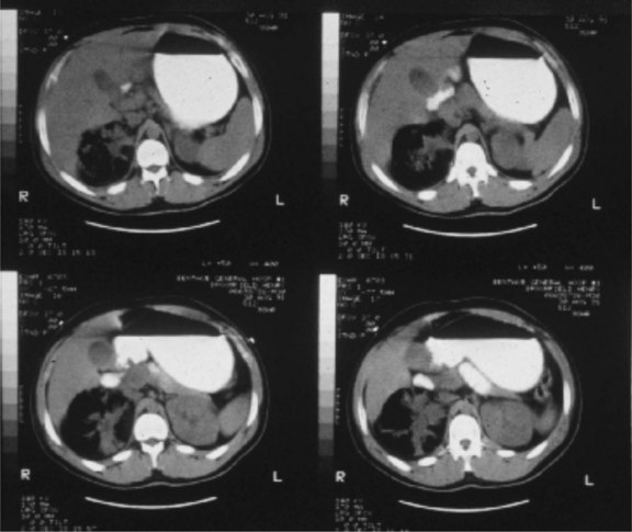
Gas-forming or emphysematous infection of the urinary tract is usually seen in the poorly controlled diabetic patient. Emphysematous pyelonephritis typically presents as a fulminant infection involving the renal parenchyma, which may progress to involve the perinephric space7 (Fig. 73-1). A lesser form of this process, emphysematous pyelitis, results in gas within the renal collecting system but not within the parenchyma. The causative agent most commonly is E. coli, which produces gas through a facultative anaerobic or fermentative process. Significant soft tissue destruction may result and a picture consistent with urosepsis is common. Control of the metabolic abnormalities and aggressive antibiotic and supportive therapy in a critical care setting are essential. If there is upper tract obstruction present, it should be relieved promptly with retrograde or antegrade tube drainage. If discrete purulent collections are present based on computed tomography (CT) imaging, percutaneous drainage may be attempted while the sepsis is being controlled medically. These patients often require urgent nephrectomy, although if the overall clinical status is improving with medical treatment, there may be an advantage to delaying surgical intervention until the patient is more stable. At times, the process may be focal or segmental and may respond solely to medical therapy and indicated drainage procedures.


Emphysematous cystitis is a gas-forming infection involving the bladder wall, with gas evident in the submucosa and/or detrusor level, also best demonstrated on a CT scan. Therapy consists of urinary catheter drainage, aggressive antibiotic therapy, and supportive care.


Acute papillary necrosis is also most commonly seen in the diabetic patient.8 Often, there is an underlying ischemic state involving the renal papillae, which progresses to frank necrosis and passage of the sloughed papilla into the collecting system and ureter, causing obstruction and, if concomitant infection is present, often progresses to urosepsis. These patients require urgent drainage of the obstructed upper tract and, after their infection is resolved, endoscopic evacuation of the necrotic tissue. This process may present as a fulminant infectious illness or as a chronically progressive process.


Gas may be present in the urinary tract from anaerobic urinary infection in the absence of these emphysematous processes. Gas may also be present because of instrumentation or catheterization or the presence of urinary tract fistulae; the most common is the colovesical fistula secondary to complications of diverticulitis or colonic neoplasms.


Xanthogranulomatous Pyelonephritis


Xanthogranulomatous pyelonephritis (XGP) is a specific clinical and histologic entity that involves the presence of a foamy, lipid-laden, macrophage infiltrate in the renal parenchyma, with extensive inflammation, fibrosis, and loss of function. It is thought to result from chronic bacterial infection, usually in the presence of stones and chronic obstruction.9 The kidney is typically nonfunctional or poorly functioning and may be a source of chronic disease, pain, persistent infection, and sometimes fistulization to the flank or adjacent organs. Nephrectomy is usually indicated. Radiographically, by CT, there may be an apparent collecting system dilation; however, drainage attempts often are unproductive because the material is often solid or too viscous to drain. Patients may present with active infection and require a cooling off period with antibiotics and supportive care to be readied for surgery. The general surgeon may become involved in such cases because of this lesion’s propensity to become densely adherent to surrounding structures, particularly the diaphragm, pancreas, duodenum, great vessels, iliac vessels, and flank wall. The risk of iatrogenic adjacent organ injury is high in these nephrectomies. Often, the renal hilum is so inflamed and fibrotic that the renal vessels cannot be individually dissected; in these cases, placement of a vascular pedicle clamp with renal excision and oversewing the pedicle is necessary.


Epididymitis, Epididymo-Orchitis, Without and With abscess


The topic of the acute scrotum is important to urologists and general surgeons because the differential diagnosis can be challenging and an accurate diagnosis may only be achievable surgically.10 The epididymis, located posterolateral to the testis, becomes infected through ascending infection from the urinary tract down the vas deferens into the scrotum. When infection is advanced, the entire scrotal contents ipsilaterally become involved, with overlying skin fixation and edema. It may be difficult to distinguish this entity from late torsion, incarcerated inguinal hernia, or testicular tumor with necrosis and inflammation. Scrotal ultrasound is useful diagnostically, especially to rule out associated abscess. Cultures should be obtained and patients should be treated with broad-spectrum antibiotics. If abscess is present, surgical drainage and often orchiectomy is indicated. If improvement is seen with medical therapy, rest, and scrotal elevation, continued observation may result in eventual, although often slow, resolution with medical therapy alone. If there is persistent pain and mass, or signs of testicular ischemia are noted by repeat Doppler imaging, exploration and orchiectomy may still be necessary to resolve the process.


Fournier’s Gangrene


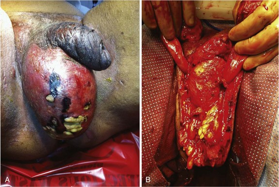
Gas-forming and necrotizing soft tissue infections are covered in detail elsewhere in this text. When the genitalia are involved, patients typically present with significant pain and tenderness, scrotal and genital swelling, discoloration or frank necrosis, crepitus and, at times, foul-smelling discharge (Fig. 73-2). Bacteriologically, these infections are usually polymicrobial, with aerobes, anaerobes, gram-positive, and gram-negative organisms. Necrotizing soft tissue infections of the genitalia, as in other anatomic regions, require aggressive broad-spectrum antibiotic treatment, supportive care, and urgent surgical drainage with aggressive débridement of the necrotic tissue.11 The magnitude of the débridement depends entirely on the degree of progression of the process. It is rare for the process to involve the testicles or deep tissues of the penis, so these structures should be preserved, if possible. I prefer to separate the parietal tunica vaginalis of the testes from the overlying necrotic dartos and skin and preserve the tunical compartment intact, which helps with wound care and patient comfort. If the penile skin is necrotic, it can be débrided down to but not through the Buck’s fascial layer. It is uncommon for the urethra to be involved although, occasionally, a defined urinary tract source may be evident, such as a urethral stricture with perforation and local infection. Suprapubic tube diversion is generally not necessary; urethral catheter drainage is generally sufficient. Wound vacuum-assisted closure (VAC) device placement after purulence has resolved results in rapid wound size reduction and fluid evacuation. Grafting large defects with meshed split-thickness grafts for the scrotum and nonmeshed thick split-thickness grafts for the penile shaft yields favorable results.


Genitourinary Fungal and Tuberculous Infections


Fungal infections of the urinary tract are most often seen in diabetics, immunocompromised patients, and those who have had extensive nosocomial and antibiotic exposure. These infections vary markedly, from superficial candidal infections of the groins and genitalia, treatable with standard topical agents, to invasive fungal infections of the bladder or kidneys that may cause urosepsis and be life-threatening.12 Fungal infections are addressed in general terms elsewhere in this text. Specific issues related to the urinary tract include the occasional need for antifungal bladder irrigation, potential for fungal deposits (fungus balls) forming in the renal colleting system requiring direct irrigation or occasionally endoscopic removal, and complex cutaneous involvement of the genitalia. Infectious disease consultation and support are valuable in such cases because the organisms may be resistant and atypical, and selection of treatment agents may not be straightforward.


Genitourinary (GU) tuberculosis may be manifested as an isolated GU infection (e.g., tuberculous cystitis, epididymitis) or as part of a systemic infection.13 Urine cultures from the first morning void are most effective in detecting infection. Current references should be consulted to address specific anti-infective therapy agent selection. Upper urinary tract tuberculosis infection may cause ureteral strictures, which may progress even with therapy and result in silent obstruction and renal loss if not promptly detected. These patients should be regularly monitored by ultrasound to assure adequate upper tract drainage. Tuberculous epididymitis should be suspected if chronic epididymitis results in cutaneous fistula formation; following adequate medical therapy, epididymectomy or orchiectomy may be indicated for residual mass, pain, or fistula. Renal nonfunction following diagnosis and treatment may occasionally require partial or total nephrectomy. As a general principle, atypical infections of the genitourinary system should prompt testing for an immunocompromised state, including testing for HIV status, because varied urologic manifestations of such viral infections may be observed.14


Voiding Dysfunction, Bladder Outlet Obstruction, Benign Prostatic Hyperplasia, And Incontinence


The accurate diagnosis and tailored management of voiding dysfunction is an important aspect of general urologic care. From simple postoperative urinary retention caused by immobility, local pain, and medication side effects, to female or male stress incontinence following pelvic surgery, to the complex neurourologic disorders of the spinal cord injury patient, voiding dysfunction is one of the most common reasons to consult an urologist. To focus on the most important and common entities in this broad range of pathology, I will discuss four examples, as well as male voiding dysfunction caused by benign prostatic hyperplasia (BPH).


Urinary Incontinence


Incontinence, or the inability to exercise full volitional control of urine passage, is often classified symptomatically. Urgency incontinence is loss of urine associated with an urge to void. Stress incontinence is loss of urine with movement, straining, or increase in abdominal pressure. Overflow incontinence is loss of urine when the bladder becomes full and there is an inability to empty volitionally. Mixed incontinence combines these.


Urge incontinence is most commonly caused by overactive bladder or detrusor instability. This may be age-related, caused by a specific anatomic bladder irritative focus, or be neurologic in origin. Urologic consultation may be necessary to determine the cause; anticholinergic or antimuscarinic medication therapy is the mainstay of treatment once critical underlying issues (e.g., bladder tumor, carcinoma in situ, obstruction) are excluded. There is a burgeoning variety of overactive bladder medications on the market. They generally have predictable side effects, including dry mouth, constipation, and often confusion in older adults, and should be avoided in patients with a history of narrow-angle glaucoma; an ophthalmologist’s approval should be sought for these patients. These drugs will decrease urgency and episodes of urge incontinence in most patients and are typically safe for long-term use. Refractory cases may require more complex interventions, including nerve stimulator devices, intravesical botulinum toxin injections and, rarely, surgical bladder augmentation.15


Stress incontinence results from anatomic changes in the pelvic floor or sphincteric apparatus that lead to excessive bladder hypermobility or sphincteric incompetence, so that with an increase in abdominal pressure, urine is involuntarily lost. Common clinical settings would be following multiple vaginal deliveries, with stretching of the pelvic floor supports and bladder prolapse, and male incontinence after radical prostatectomy. The treatment may be behavioral (e.g., biofeedback training, pelvic floor exercises), involve injectable bulking agents (injected into the sphincteric region) or surgical (e.g., female pelvic floor reconstruction, female or male sling device implants, artificial urinary sphincter device implant). This is a specialized area of urologic care and surgery. Urodynamics testing may be useful in assessing the sphincter anatomy and function prior to initiating therapy.


Overflow incontinence is involuntary loss of urine caused by excessive bladder fullness. It is often missed as a diagnosis; careful history, physical examination to palpate or percuss the full bladder, and/or measurement of postvoid residual by ultrasound or catheter drainage are essential. Treatment is based on determining the cause of the bladder distention (obstructive versus detrusor dysfunction) and managing that issue accordingly—for example, by corrective surgery for bladder outlet obstruction or self-intermittent catheterization for refractory detrusor failure.


It is common for incontinence to be mixed—that is, to have elements of stress and urgency. Urodynamic testing is important in such cases to distinguish obstruction from detrusor dysfunction and to plan therapy.16 In general, the medical component (e.g., bladder control medicine for urgency) is treated first, with surgical intervention reserved for patients in whom medical management is not effective or does not address a critical underlying problem, such as high-grade outlet obstruction.


Benign Prostatic Hyperplasia and Bladder Outlet and Urethral Obstruction


BPH is generally considered to be the most common benign internal neoplasm of the adult male. It is an almost ubiquitous process in men, although varying greatly in degree, bothersomeness, and complications.


BPH may result in LUTS (lower urinary tract symptoms) and BOO (bladder outlet obstruction). The process involves hypertrophy and hyperplasia, with increased glandular and stromal elements of the prostate in varying amounts. There is little correlation between the measured volume of the prostate and degree of symptomatology that results. In addition, the degree of BOO does not necessarily correlate with the severity of LUTS. Symptoms may be mild and manageable just with watchful waiting, or may be more significant, requiring long-term medical therapy and, at times, surgical intervention. Complications of BPH may include bladder damage and loss of function from chronic distention, upper tract deterioration, troublesome hematuria, bladder stone formation, and recurrent urinary infection. Practice guidelines for BPH have been produced by the American Urologic Association to guide providers in the diagnosis and management of BPH.17 The basic evaluation involves history and physical examination, digital rectal examination, urinalysis, prostate-specific antigen (PSA) level testing (in select patients), other interventions to rule out prostate cancer and, at times, other tests to rule out other significant urologic pathology (e.g., urine cytology, upper tract imaging, cystoscopy). A quantitative symptom score may be useful. Checking PVR urine volume may be valuable as well. After the basic assessment, patients are directed toward some therapeutic approach, such as watchful waiting, medical therapy, or minimally invasive or standard surgical intervention. The management selection reflects patient preferences and the presence of complicating factors.


Medical therapy options consist of α-adrenergic blocking agents and/or 5-alpha-reductase inhibitors. The former work on the α or α1a receptors (e.g., tamsulosin) of the bladder outlet, relaxing smooth muscle in the prostatic stroma and bladder neck region. They may cause some orthostatic side effects, which are usually mild. 5-Alpha-reductase inhibitors (e.g., finasteride, dutasteride) block the conversion of testosterone to dihydrotestosterone, the active agent that causes and maintains BPH. These agents will reduce the actual volume of the prostate, with maximal effects seen by 6 months, and maintain that volume reduction during continued use. This class of drugs also alters the serum PSA level (reduces it ≈50%), which must be kept in mind with regard to prostate cancer screening.


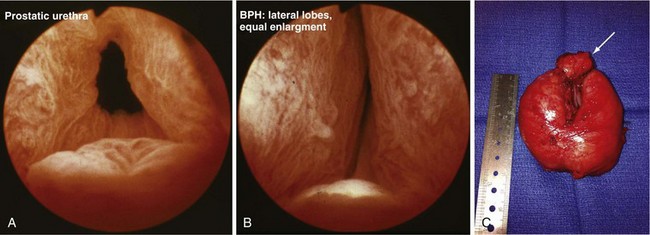
When medical therapy is ineffective and symptoms remain bothersome, or when there is an objective surgical indication (e.g., severe obstruction based on urodynamic data, recurrent hematuria or infection, urinary tract anatomic deterioration from obstruction), surgical intervention is considered. The standard approaches include minimally invasive options (e.g., laser procedures, thermotherapy, microwave procedure), TURP or, when the adenomatous growth is particularly large, open simple prostatectomy to enucleate the adenoma surgically (Fig. 73-3). Complications of these surgical procedures include persistent bleeding, prostatic capsular perforation, or perforation into periprostatic venous sinuses, with fluid absorption, which may result in hyponatremia because of the glycine irrigation typically used (newer electroresection systems use normal saline with a bipolar electrode, eliminating the hyponatremia risk) and, rarely, injury of an adjacent structure (e.g., rectal injury with TURP or open prostatectomy).


Because BOO and LUTS from BPH are so common in today’s aging population, the general surgeon will deal with many patients with this diagnosis. If any difficulty is encountered when catheterizing a patient with BPH, a coude-tipped catheter should be used, with the angulated tip oriented cephalad; this will allow atraumatic catheterization for many BPH patients. If this is not successful, a urologist should be consulted to assess the patient and determine whether there is another cause of obstruction, such as a urethral stricture or bladder neck contracture, which may be present in up to 10% of patients post-TURP; this may require a more specialized technique for urethral passage.


Male Reproductive And Sexual Dysfunction


An area of practice to which urologists devote significant attention is male infertility and sexual dysfunction.18 Diagnostic evaluation, medical treatment, and surgical therapy represent sophisticated aspects of urologic care; general surgeons should have a basic familiarity with the management of these disorders. They may at times be called on to participate in the treatment of surgical complications of genital surgery and prosthetic implant surgery; the patient’s history of having undergone these procedures may affect the precautions and challenges of nonurologic pelvic and groin procedures.


Male Infertility: Evaluation and Treatment


Infertility affects approximately 15% to 20% of couples; the male factor is primary or sole factor in 50% of these cases. Couples are often referred to the urologist following a period of infertility, and referrals are generally from a primary care physician and often from the evaluating gynecologist. It is not uncommon in my experience as a urologist to receive a patient following an exhaustive workup and extensive treatment for female reproductive dysfunction only to discover late in the process that there is a significant male factor, typically identified by semen analysis.


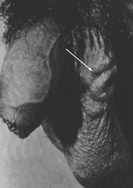
The standard male factor evaluation involves a detailed history, physical examination, and basic laboratory and imaging evaluation. The history should include a discussion regarding sexual and reproductive history, including potential gonadotoxic exposure, urologic and sexually transmitted infections, trauma and prior surgery involving the pelvis, groin, and genitalia, and family history of infertility. Physical assessment should include a general evaluation of masculinization, genital findings, including normal meatal location, testicular size and, consistently, presence and normalcy of the epididymis and vas deferens, and possible presence of a varicocele (Fig. 73-4). Perineal and rectal examinations are routine parts of this assessment.


Basic Laboratory Assessment


Laboratory evaluation includes a semen analysis and serum hormone studies. Semen analysis parameters of importance include semen volume and consistency, sperm concentration and total count, percentage motility and quality of sperm movement, sperm morphology, and presence of red and white blood cells or bacteria. The World Health Organization has defined parameters of normal for routine semen analyses.


Semen analysis abnormalities fall into two main categories—azoospermia (the complete absence of sperm from the semen), and abnormal bulk semen parameters (e.g., reduced concentration, motility, morphology [in isolation or in combination], and abnormal sperm function). Azoospermia results from ablative pathology of the germinal epithelium of the testis with absence of the production of mature sperm or from defects in sperm transport or ejaculation, often caused by ductal obstruction or ejaculatory dysfunction. When azoospermia is caused by lack of sperm production, this is often accompanied by normal semen volume and by a markedly elevated serum follicle-stimulating hormone (FSH) level. There are exceptions to this rule; however, because certain forms of maturational arrest of intratesticular sperm development, as well as other sperm production defects, can be seen with normal serum hormone parameters. Abnormalities of sperm transport are commonly caused by epididymal obstruction or obstruction at the level of the ejaculatory duct within the prostate, or may be caused by remote iatrogenic injury (e.g., vasal occlusion in the groin from pediatric inguinal hernia repair, and adult hernia repair with vasal entrapment from permanent polypropylene mesh). Abnormal bulk semen parameters may be indicative of a wide range of disorders that go beyond the scope of this discussion; important entities that may cause reduced sperm numbers, motility, or morphology include varicocele, antisperm antibodies causing immunologic infertility, genital duct infection with pyospermia causing sperm dysfunction, and prior or current gonadotoxic exposure. Reduced semen volume may be artifactual, indicating incomplete ejaculation or specimen collection, or may represent true pathology, including, for example, congenital absence of the seminal vesical, ejaculatory duct obstruction, or retrograde ejaculation caused by diabetes or neurologic injury or prior bladder neck surgery or medications.


Serum hormone testing includes determining levels of FSH, luteinizing hormone (LH), testosterone, free testosterone (measured or calculated based on binding protein levels), and prolactin. It may be possible to diagnose hypogonadotropic hypogonadism on the basis of serum hormone studies or note significant elevation in the FSH level, which is often indicative of significant ablative intratesticular pathology. In any situation in which the serum testosterone level is below normal, the serum prolactin level should be measured to rule out a prolactinoma, a benign but potentially clinically significant neoplasm of the pituitary gland.


Additional testing of value in the evaluation of the infertile male may include ultrasound imaging and additional sperm function or genetic testing. Ultrasound of the scrotum is useful to measure testicular volume and symmetry, exclude the possibility of testicular neoplasm, identify epididymal anatomy, and define or confirm the presence of a varicocele, which is an abnormal dilation of the pampiniform venous plexus of the internal spermatic venous system. This may cause testicular overheating, reduction in the level and quality of sperm production, testicular atrophy, and discomfort or pain. Transrectal ultrasound of the prostate may provide evidence of ejaculatory duct obstruction with seminal vesical dilation or congenital absence of the seminal vesicle, which may accompany congenital absence of the vas deferens.


Additional laboratory testing may include ultrastructural studies of sperm for motility analysis, semen leukocyte assay, antisperm antibody assay, and biologic tests of sperm-egg interaction; these more advanced tests have value in pursute of a diagnosis in cases of unexplained infertility. Patients with severe oligospermia or azoospermia should have genetic testing performed to rule out a significant genetic cause of the infertility; this information may affect a couple’s decision about whether and how to proceed with fertility treatment attempts. Screening for cystic fibrosis mutations, Y chromosome microdeletion assays, sperm chromatin damage assays, and basic karyotype analysis are typically included in this genetic evaluation.


Treatment of male infertility depends on the identified cause and on the availability and affordability of assisted reproductive technologies support options for specific or empirical treatment of failure to conceive. Commonly used medical therapies include hormonal stimulation of spermatogenesis, which has had mixed success, and anti-inflammatory or antibiotic therapy for pyospermia or genital duct infection. Surgical therapies may include microsurgical reconstruction for vasal or epididymal occlusion (including vasectomy reversal), transurethral resection of the ejaculatory duct for obstructive lesions, and varicocele repair, which may be performed by open groin surgery approach, laparoscopically, or through interventional radiology internal spermatic vein embolization.


Surgeons must be aware of the potential for groin and pelvic surgical procedures to cause male infertility via damage to the spermatic cord vasculature, vas deferens, or ejaculatory duct region, or from vasal entrapment from mesh used for inguinal hernia repair. The vas and testicular blood supply is vulnerable to injury, particularly when the groin is explored in a reoperative setting for recurrent inguinal hernia or when the anatomy is obscure because of trauma, because the specific identification of these structures may be challenging. If the vas is injured during a general surgical procedure and the patient is in his reproductive years, intraoperative urologic consultation may be appropriate.

< div class='tao-gold-member'>

Stay updated, free articles. Join our Telegram channel

Aug 1, 2016 | Posted by in CARDIAC SURGERY | Comments Off on Urologic Surgery

Full access? Get Clinical Tree

Get Clinical Tree app for offline access