Unstable Angina/Non-ST-Elevation MI


Unstable Angina/Non-ST-Elevation MI


Stanley Chetcuti

Eric R. Bates


Unstable angina (UA) and non-ST-segment elevation myocardial infarction (NSTEMI) form part of the spectrum of acute coronary syndromes (ACSs) that includes acute ST-segment-elevation myocardial infarction (MI) and sudden cardiac death. Both conditions are pathophysiologically and clinically related and may be indistinguishable at the time of presentation. Patients with biochemical marker evidence for myocardial necrosis are ultimately defined as having NSTEMI instead of UA. The causes of UA and NSTEMI may be classified under five different headings (Table 10.1).


USUAL CAUSES

The usual cause of UA/NSTEMI is disruption of an atherosclerotic plaque and formation of a nonocclusive thrombus. Plaques prone to rupture have a large lipid core, high macrophage and activated T-lymphocyte density, low smooth muscle cell density, and a thin fibrous cap characterized by disorganized collagen (1,2). The plaque shoulder, at its junction with the arterial wall, is mechanically the weakest point where most ruptures occur, exposing the lipid core that is very potent in stimulating platelet-rich thrombus formation (3,4). Two-thirds of plaque ruptures with thrombus have a less than 50%-diameter stenosis before plaque rupture and 97% have less than 70%-diameter stenosis (5). Thrombus occurring on a ruptured or fissured plaque results from a complex series of interactions between the exposed lipid core, macrophages, smooth muscle cells, collagen, circulating blood products, and coagulation factors. Platelet surface receptors recognize the vascular matrix components (collagen, von Willebrand factor, vitronectin, and fibronectin), stimulating platelet activation and adhesion. Activated platelets secrete mitogenic, chemotactic, and vasoactive substances and undergo conformational changes with the recruitment and activation of glycoprotein (GP) IIb/IIIa receptors. The activated GP IIb/IIIa receptors mediate platelet aggregation by fibrinogen cross-linkage, forming the white thrombus on the surface of the plaque (6). Tissue factor interacts with activated factor VII to initiate the coagulation cascade, resulting in the generation of fibrin, which traps red blood cells and forms the overlying red thrombus (7). Myocyte necrosis in NSTEMI is believed to be due to temporary arterial occlusion or embolization of platelet-thrombus aggregates and plaque material into the microcirculation.

Less-common causes include dynamic obstruction, progressive atherosclerosis or restenosis, and inflammation. Vasoconstrictor substances acting on a segment of epicardial coronary artery with dysfunctional endothelium may lead to vasoconstriction or focal spasm (8,9). Progressive atherosclerotic obstruction can occur in stable calcified lesions or after percutaneous coronary intervention (PCI). Sites of plaque disruption usually exhibit features of inflammation (10). It is not completely clear what role certain infectious agents have on inflammation, but
they may be related to increased concentrations of activated macrophages and T lymphocytes at the plaque shoulder.


TABLE 10.1. Causes of unstable angina and non-Q-wave MI


1.


Nonocclusive thrombus on pre-existing plaque


2.


Dynamic obstruction (coronary artery spasm or vasoconstriction)


3.


Progressive mechanical obstruction


4.


Inflammation and/or infection


5.


Secondary unstable angina


These causes are not mutually exclusive; some patients have two or more causes.


Reprinted with permission from Braunwald E. Unstable angina: an etiologic approach to management. Circulation 1998;98:2219-2222.


Noncardiac events can cause a mismatch in myocardial oxygen demand and supply, resulting in UA/NSTEMI. They may be caused by (a) increased myocardial oxygen demand (fever, thyrotoxicosis), (b) reduced myocardial oxygen delivery (anemia, hypoxemia), or (c) reduced coronary blood flow (arrhythmia, hypotension). Although coex-isting coronary artery disease may be present, it is usually stable, and care should be focused on the precipitating condition.


PRESENTING SYMPTOMS AND SIGNS

The main complaint of patients with UA and NSTEMI is worsening, or new-onset, angina. Three principal presentations of unstable angina are seen (11) (Table 10.2). The character of the angina is the same as that encountered in chronic stable angina, but is usually more severe and of longer duration, may occur at rest, or may be precipitated by less exertion than previously.

Classically, the pain is described as a pressure sensation in the chest that often radiates to the left arm and neck. Associated with the chest pain in varying frequencies are the symptoms of diaphoresis, dyspnea, nausea, and vomiting. Occasionally, patients (especially elderly women) may have no discernable chest pain but may complain of varying components of arm pain, neck pain, and epigastric discomfort. A decrease in exercise threshold with worsening dyspnea may be noted on exertion. When these non-chest pain symptoms are clearly related to physical or emotional stress and are relieved by nitroglycerin, they should be considered anginal equivalents. Progression in frequency and intensity should warrant the same degree of concern as with chest pain.


TABLE 10.2. Three principal presentations of unstable angina


Rest angina


Angina occurring at rest and prolonged, usually >20 min


New-onset angina


New-onset angina of at least CCS class III severity


Increasing angina


Previously diagnosed angina that has become distinctly more frequent, longer in duration, or lower in threshold (i.e., increased by one or more CCS class to at least CCS class III severity)


CCS, Canadian Cardiac Society Classification.


Reprinted with permission from Braunwald E. Unstable angina: a classification. Circulation 1989;80:410-414.


A careful history may unmask features in the chest-pain syndrome that make it more likely to be noncardiac in origin. Pain of extreme duration, that lasts for many hours or days, or at the other end of the spectrum, a few seconds, is less likely to be ischemic in origin. Pain that is clearly pleuritic or positional or located with the tip of one finger is also unlikely to be cardiac in origin. The physician should document in the medical record whether a high, intermediate, or low likelihood exists of acute ischemia caused by coronary artery disease (Table 10.3).

The physical examination is often unremarkable, with no abnormal findings of the pulse, blood pressure, chest, or cardiovascular system. It is very important that the examination be performed in a meticulous and structured fashion, because it may elucidate noncardiac causes of chest pain (pleurisy, pneumothorax) and nonischemic causes of cardiac pain (valvular disease, pericarditis,
and vascular emergencies). Serial examinations may uncover signs such as hypotension, brady/tachycardia, pulmonary rales, a third heart sound, or a new or worsening murmur that predict higher risk and demand a more aggressive management (diagnostic and treatment) pathway (12).


TABLE 10.3. Likelihood that signs and symptoms represent an ACS secondary to CAD


FEATURE


HIGH LIKELIHOOD:
ANY OF THE FOLLOWING


INTERMEDIATE LIKELIHOOD:
ABSENCE OF HIGH-
LIKELIHOOD FEATURES
AND PRESENCE OF ANY
OF THE FOLLOWING


LOW LIKELIHOOD:
ABSENCE OF HIGH- OR
INTERMEDIATE-
LIKELIHOOD FEATURES
BUT MAY HAVE


History


Chest or left-arm pain or discomfort as chief symptom reproducing prior documented angina


Chest or left-arm pain or discomfort as chief symptom


Age >70 yr


Probable ischemic symptoms in absence of any of the intermediate-likelihood characteristics


Known history of CAD, including MI


Male sex


Diabetes mellitus


Recent cocaine use


Examination


Transient MR, hypotension, diaphoresis, pulmonary edema, or rales


Extracardiac vascular disease


Chest discomfort reproduced by palpation


ECG


New, or presumably new, transient ST-segment deviation (≥0.5 mm) or T-wave inversion (≥2 mm) with symptoms


Fixed Q waves


Abnormal ST segments or T waves not documented to be new


T-Wave flattening or inversion in leads with dominant R waves


Normal ECG


Cardiac markers


Elevated cardiac TnI, TnT, or CK-MB


Normal


Normal


ACS, acute coronary syndrome; CAD, coronary artery disease; CK-MB, creatine kinase MB, ECG, electrocardiogram; MI, myocardial infarction; MR, mitral regurgitation; TnI, troponin I; TnT, troponin T.


Reprinted with permission from Galvani M, Ottani F, Oltrona L, et al. N-terminal pro-brain natriuretic peptide on admission has prognostic value across the whole spectrum of acute coronary syndromes. Circulation 2004;110:128-134.


HELPFUL TESTS


Electrocardiography

The ECG is important for both diagnostic and risk-stratification purposes. Specific characteristics and the magnitude of pattern abnormalities increase the likelihood of coronary artery disease. New or dynamic ST-segment depression (>0.5 mm) is suggestive of acute ischemia with an associated prothrombotic state (13). Inverted T waves may also suggest ischemia or NSTEMI, although the risk is less than for ST-segment depression. Nonspecific ST-segment changes (0.5 mm or less) and T-wave changes (2 mm or less) are much less helpful and may also be related to drugs (phenothiazines, digitalis, etc.), hyperventilation, or repolarization abnormalities in association with left ventricular hypertrophy or conduction disturbances. Conversely, the ECG may be normal in 1% to 6% of patients with NSTEMI and more than 4% of patients with unstable angina (14).

In the GUSTO-IIb trial, the 30-day incidence of death or myocardial infarction was 5.5% in patients with T-wave inversion, 9.4% in those with ST-segment elevation, 10.5% in those with ST-segment depression, and 12.4% in patients with ST elevation and
depression (15). All these ECG findings may be transient phenomena, and this illustrates the importance of serial tracings. Continous ST-segment monitoring may reveal episodes of otherwise undiagnosed ischemic episodes.


Biochemical Markers

Although many markers and assays that detect myocardial necrosis are available, the cardiac troponins T and I and the creatinine kinase MB (CK-MB) isoform are the most commonly used, with the the troponins gaining acceptance as the markers of choice in acute coronary syndromes. They have achieved an important role in diagnostic, prognostic, and treatment pathways by virtue of their high degree of sensitivity and specificity and their relative ease of use and interpretation. The joint statement of the European Society of Cardiology and the American College of Cardiology declared that myonecrosis may be diagnosed when the maximal concentration of troponin T or I exceeds the decision limit (99th percentile for a reference group) on at least one occasion in a 24-hour period (16). This new definition has increased the frequency of the NSTEMI diagnosis in acute coronary syndrome patients by 30%. Troponin I is more accurate with renal insufficiency. The troponins are both detectable about 6 hours after myocardial injury and are measurable for up to 2 weeks. Mortality risk is directly proportional to troponin levels, and the prognostic information is independent of other clinical and electrocardiographic risk factors (17,18) (Fig. 10.1).


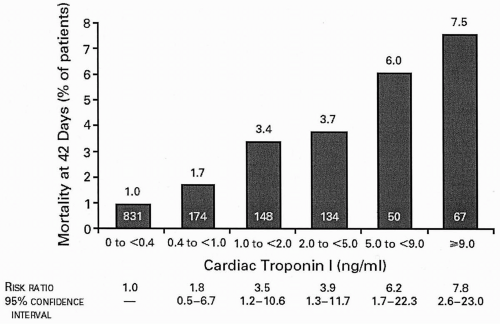
FIGURE 10.1. Mortality rates at 42 days according to the level of cardiac troponin I measured at enrollment. (Adapted from Antman EM, Tanasijevic MJ, Thompson B, et al. Cardiac-specific troponin I levels to predict the risk of mortality in patients with acute coronary syndromes. N Engl J Med 1996;335:1342-1349, with permission.)

CK-MB is less specific, also being present in skeletal muscle and in low levels in the blood of healthy persons. Unlike troponins, it is useful in detected recurrent myocardial necrosis early after the initial event, as levels tend to return to normal within 36 to 48 hours after initial release.

The role of increased inflammatory activity as an independent risk marker in
acute coronary syndromes (ACSs) continues to elicit vigorous research interest. C-reactive protein (CRP) levels in ACS appear to be related to long-term mortality in an independent and additive fashion to troponin levels (19). The role of other inflammatory markers, such as amyloid A and interleukin-6, in the day-to-day management of patients with ACS, is as yet undefined.

Elevated levels of fibrinogen, fibrinopeptide-A, and plasminogen activator inhibitor-1 (PAI-1) have all been shown, albeit not consistently, to be related to an increased and independent risk of long-term events in patients with ACS (20,21,22). Again, these markers are not yet routinely used in clinical practice, as their additive role is not yet defined.

B-Type natriuretic peptide (BNP) is a neurohormone released in its precurser form, proBNP, when the ventricular myocardium is stretched. BNP and its precurser can be measured easily in many hospital laboratories. Increasing serum levels of these neurohormones measured at first contact or early in the hospital stay are associated with higher short- and long-term mortality rates (23).


TABLE 10.4. Noninvasive risk stratification


HIGH RISK (>3% ANNUAL MORTALITY RATE)


1.


Severe resting LV dysfunction (LVEF <35%)


2.


High-risk treadmill score (score ≤-11)


3.


Severe exercise LV dysfunction (exercise LVEF <35%)


4.


Stress-induced large perfusion defect (particularly if anterior)


5.


Stress-induced multiple perfusion defects of moderate size


6.


Large, fixed perfusion defect with LV dilation or increased lung uptake (thallium 201)


7.


Stress-induced moderate perfusion defect with LV dilation or increased lung uptake (thallium 201)


8.


Echocardiographic wall-motion abnormality (involving more than two segments) developing at a low dose of dobutamine (≤10 mcg/kg/min) or at a low heart rate (<120 beats/min)


9.


Stress echocardiographic evidence of extensive ischemia


INTERMEDIATE RISK (1% TO 3% ANNUAL MORTALITY RATE)


1.


Mild/moderate resting LV dysfunction (LVEF 35%-49%)


2.


Intermediate-risk treadmill score (-11 < score <5)


3.


Stress-induced moderate perfusion defect without LV dilation or increased lung intake (thallium 201)


4.


Limited stress echocardiographic ischemia with a wall-motion abnormality only at higher doses of dobutamine involving two or more segments


LOW RISK (<1% ANNUAL MORTALITY RATE)


1.


Low-risk treadmill score (score ≥5)


2.


Normal or small myocardial perfusion defect at rest or with stress


3.


Normal stress echocardiographic wall motion or no change of limited resting wall-motion abnormalities during stress


LV, left ventricular; LVEF, left ventricular ejection fraction.


Reprinted with permission from Fuster V, Lewis A. Conner Memorial Lecture: Mechanisms leading to myocardial infarction: insights from studies of vascular biology. Circulation 1994;90:2126-2146.


Noninvasive Testing

Echocardiography allows the rapid determination of left ventricular function. Stress testing to risk stratify (Table 10.4) should be performed in low-risk and intermediate-risk patients free of complications who are not referred for coronary angiography (24). Choice of stress test is based on the resting ECG, ability to exercise, and local expertise. Treadmill testing is suitable in patients with good exercise tolerance in whom the ECG is free of ST-segment abnormalities, bundle-branch block, left ventricular hypertophy,
intraventricular conduction delay, paced rhythm, preexcitation, and digoxin effect. Echocardiographic or nuclear stress imaging should be added in patients with ECG abnormalities that prevent accurate interpretation. Pharmacologic stress testing can be performed in patients who cannot achieve an adequate exercise stress on the treadmill.


Cardiac Catheterization

Cardiac catheterization defines regional and global left ventricular function, valvular function, and coronary artery anatomy. It is routinely performed 1 to 2 days after hospital admission in patients treated with the “early invasive strategy,” which is directed toward immediate coronary revascularization. Alternatively, the “early conservative strategy” involves reserving cardiac catheterization for patients with recurrent angina receiving aggressive medical therapy or for those with ischemic stress-test results. The test should not be performed in patients who are clearly not revascularization candidates, in those who do not want catheterization, or in those who are at low risk. Indications for cardiac catheterization are listed in Table 10.5 (24).


COMPLICATIONS

If left untreated, 5% to 10% of patients with UA die, and 10% to 20% have nonfatal MI within 30 days. In one-fourth of patients with NSTEMI, Q-wave MI develops, with the remainder having non-Q-wave MI. Arrhythmia, congestive heart failure, and cardiogenic shock are life-threatening complications. Recurrent ischemia may result in the need for urgent coronary artery revascularization. The GRACE Prediction score card (25) (Fig. 10.2) and the TIMI Risk Score (Fig. 10.3) (26) are prognostic tools that have been shown to predict death, MI, and need for urgent revascularization.


DIFFERENTIAL DIAGNOSIS

Chest pain, the main manifestation of ACS, may be the presentation of many nonischemic conditions. The rapid evaluation and treatment that is warranted for ACS should not be done at the expense of potentially missing an alternative condition that would warrant a significantly different approach and treatment.

Causes of nonischemic chest discomfort include the following: (a) musculoskeletal chest pain; (b) gastrointestinal discomfort (gastroesophageal reflux disease, peptic ulcer disease, biliary or pancreatic disease, or esophageal spasm); (c) cardiac nonischemic pain (valvular heart disease, hypertrophic cardiomyopathy, pulmonary hypertension, pericarditis); (d) pulmonary discomfort (pulmonary embolus, pneumothorax, pneumonia, COPD exacerbation); and (e) anxiety. This list is not meant to be exhaustive, but demonstrates the spectrum of conditions and underscores the importance of a rapid and accurate diagnosis (see Chapter 1).


THERAPY

UA results in more than 2 million hospital admissions per year, and NSTEMI accounts for more than 60% of all MI admissions. The goals of an effective treatment strategy should be relief of ischemia and prevention of the serious adverse outcomes of MI/recurrent MI and death. These goals may be achieved by the initiation of appropriate therapy (Table 10.6), ongoing risk stratification, and selective coronary artery revascularization.

Only gold members can continue reading. Log In or Register to continue

Stay updated, free articles. Join our Telegram channel

Aug 18, 2016 | Posted by in CARDIOLOGY | Comments Off on Unstable Angina/Non-ST-Elevation MI

Full access? Get Clinical Tree

Get Clinical Tree app for offline access