Heart Failure with Preserved Systolic Function


Heart Failure with Preserved Systolic Function


Audrey H. Wu

Mauro Moscucci

Ragavendra R. Baliga


Heart failure with preserved systolic function (HF-PSF) is the clinical syndrome of heart failure—manifesting with dyspnea, effort intolerance, and edema—that occurs in the setting of a normal ejection fraction. Although the abnormality in systolic heart failure leads to the impaired ability of the ventricle to eject blood, the structural or functional abnormality in HF-PSF is the impaired ability of the ventricle to fill with blood. Pure HF-PSF excludes other causes of heart-failure symptoms that occur in the context of normal ejection fraction, including pericardial or valvular disease, which may require condition-specific treatment distinct from the general treatment principles for HF-PSF. The diagnosis of HF-PSF in many cases remains difficult, in part because of the lack of standard diagnostic criteria. In addition, compared with systolic heart failure, significantly fewer clinical trial data are available to guide the clinician on best treatment strategies for HF-PSF. Differentiation between the two entities is vital to determining appropriate treatment and prognosis. Criteria have been proposed in an attempt to diagnose this condition more accurately (1) (Table 15.1).

The first part of this chapter discusses background and general principles of management of HF-PSF, and the second part discusses aspects of specific cardiomyopathies that manifest primarily with HF-PSF.


EPIDEMIOLOGY AND USUAL CAUSES

Estimating the incidence, prevalence, and mortality—and hence, prognosis—of HF-PSF has been complicated by heterogeneous diagnostic criteria used in previous epidemiologic studies. Whereas early published reports found a wide range of prevalence (13% to 74%) and annual mortality (1.3% to 17.5%), (2) more recent studies found a prevalence ranging from one third to almost one half of community-based and hospitalized populations. (3,4,5). Exacerbations of heart failure due to HF-PSF carry the same high hospital readmission rate as do those of heart failure due to systolic dysfunction (6), and it has been estimated that HF-PSF is responsible for roughly 25% of the total health care cost of treating heart failure in the United States (7). The prevalence of HF-PSF appears to have increased over the past 15 years, although survival has not improved, in contrast with trends in survival with systolic heart failure (5). Although mortality with HF-PSF currently appears to be very similar to or slightly better than that with systolic heart failure, survival for both entities remains poor; one study observed 5-year mortality rates of 65% and 68% after heart-failure hospitalization for HF-PSF and systolic heart failure, respectively (5). Compared with individuals with systolic HF, a greater proportion of female gender, obesity, hypertension, and atrial fibrillation is found among those with HF-PSF, and patients with HF-PSF also tend to be older (4,5).

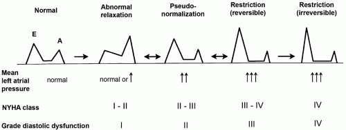
Many processes can cause the diastolicfilling abnormalities that lead to HF-PSF, although the final common pathway in pure diastolic heart failure involves impaired relaxation, decreased passive elasticity, or both. It is important to exclude other
conditions that may also present with heart failure and normal ejection fraction (Table 15.2). In primary diastolic heart failure, as a result of these myocardial compliance abnormalities, the ventricle is stiffened and requires higher pressures for filling. Consequently, the heart is particularly sensitive to alterations in factors that affect ventricular filling, including volume status, preload, afterload, and diastolic filling time. Perturbations of any of these factors can precipitate acute pulmonary edema and decompensated heart failure. Although the ejection fraction is normal, patients with HF-PSF demonstrate effort intolerance because of impaired cardiac output at rest and diminished capacity to augment cardiac output with exercise (8).


TABLE 15.1. Criteria for diagnosis of diastolic dysfunction


PROBABILITY
OF HF-PSF


DEFINITIVE EVIDENCE
OF HEART FAILUREa


EVIDENCE OF NORMAL LV SYSTOLIC FUNCTION
WITHIN 72 HR OF HEART FAILUREb


OBJECTIVE EVIDENCE OF
DIASTOLIC DYSFUNCTIONc


Definite


+


+


+


Probable


+


+


Possible


+


+


but not present at time of heart failure


LV, left ventricular.


a Clinical symptoms, signs, supporting tests such as chest radiograph, typical clinical response to treatment with diuretics, with or without documentation of elevated LV filling pressure or a low cardiac index.

b Left ventricular ejection fraction of ≥50%.

c Abnormal LV relaxation/filling/distensibility indices on cardiac catheterization or echocardiography.


From: Vasan RS, Levy D. Defining diastolic heart failure: a call for standardized diagnostic criteria. Circulation 2000;101:2118-2121, with permission.


TABLE 15.2. Differential diagnosis of heart failure with normal left ventricular ejection fraction


Incorrect diagnosis of heart failure


Inaccurate measurement of left ventricular ejection fraction


Primary valvular disease


Restrictive (infiltrative) cardiomyopathies


Amyloidosis, sarcoidosis, hemochromatosis


Pericardial constriction


Episodic or reversible left ventricular systolic dysfunction


Severe hypertension, myocardial ischemia


Heart failure associated with high metabolic demand (high-output states)


Anemia, thyrotoxicosis, arteriovenous fistulae


Chronic pulmonary disease with right heart failure


Pulmonary hypertension associated with pulmonary vascular disorders


Atrial myxoma


Diastolic dysfunction of uncertain origin


Obesity.


From Hunt SA, et al. ACC/AHA 2005 guideline update for the diagnosis and management of chronic heart failure in the adult: a report of the American College of Cardiology/American Heart Association Task Force on Practice Guidelines (Writing Committee to update the 2001 Guidelines for the Evaluation and Management of Heart Failure). J Am Coll Cardiol 2005;46:e1-82, with permission.


PRESENTING SYMPTOMS AND SIGNS

Symptoms and signs of HF-PSF are generally indistinguishable from those of heart failure due to systolic dysfunction, including dyspnea, peripheral and pulmonary edema, and exercise intolerance. The finding of elevated blood pressure during acute heart failure suggests HF-PSF as the etiology, although both the sensitivity (61% to 66%) and specificity (59% to 70%) are low. Other clinical factors that suggest HF-PSF over systolic heart failure include older age, obesity, or female gender (9,10). Differentiating HF-PSF from systolic heart failure is extremely difficult and arguably not possible with a great degree of accuracy without the assistance of imaging studies to determine ejection fraction.


HELPFUL TESTS

An assessment of cardiac function is absolutely necessary for the diagnosis of HF-PSF. Currently the optimal imaging method for this purpose is echocardiography. In addition to ejection fraction, echocardiography provides information on cardiac chamber size, wall thickness, valvular structure and function, and parameters of diastolic function. Other information that the echocardiogram may provide includes presence of pericardial effusion or thickening, estimates of central venous and pulmonary artery pressures, or abnormal myocardial echotexture, which may be found in some infiltrative cardiomyopathies. Other imaging methods used to assess ejection fraction include radionuclide studies, cardiac computed tomography, or cardiac magnetic resonance imaging. Radionuclide studies do not provide information on valvular structure and function. Cardiac computed tomography and magnetic resonance imaging, although quickly evolving and promising in providing similar structural and functional information, are not as widely available as echocardiography.

Severity and progression of HF-PSF can be assessed by examining transmitral flow velocity by Doppler echocardiography (11). Flow across the mitral valve is reflective of the relative driving force across the valve, primary contributors of which include left atrial pressure and left ventricular pressure. In turn, many factors determine left ventricular pressure, including ventricular compliance, preload, and afterload. In the early stages of diastolic dysfunction, impaired ventricular relaxation results in impaired diastolic filling (low E velocity and prolonged deceleration time) and relatively more diastolic filling with atrial contraction later in diastole (therefore low E/A ratio; grade I diastolic dysfunction). With disease progression, left atrial pressure increases and consequently driving pressure across the mitral valve increases, resulting in an increasing E velocity and shortening deceleration time (“pseudonormal” pattern, grade II diastolic dysfunction). With moreadvanced disease, a restrictive filling pattern emerges, with a high E velocity and prolonged isovolumic relaxation time (grade III to IV diastolic dysfunction). In addition to gauging disease severity, measures of diastolic function may be associated with prognosis, particularly in infiltrative cardiomyopathies (Fig. 15.1).


FIGURE 15.1. Diagram of a grading system for diastolic dysfunction, based on the progression of disease patterns in patients with cardiac disease. The schematic represents the mitral flow-velocity curve, composed of early diastolic filling (E) and atrial systole (A), in various stages of diastolic dysfunction. The natural progression is from the normal pattern to the abnormal relaxation pattern, to the pseudonormalization pattern, to a reversible restriction pattern, and finally to an irreversible restriction pattern. (Adapted from: Nishimura RA, Tajik AJ. Evaluation of Doppler filling of left ventricle in health and disease: Doppler echocardiography is the clinician’s Rosetta stone. J Am Coll Cardiol 1997;30:8-18.


Normal heart size on chest radiography is helpful in suggesting HF-PSF only when it is present; however, as left ventricular hypertrophy frequently occurs in these patients, normal heart size is not a common finding. Physical examination findings such as third or fourth heart sounds are not helpful in distinguishing HF-PSF from systolic heart failure (9). The electrocardiogram may reveal atrial fibrillation or ischemia, which can be precipitating factors to heart-failure decompensation, or evidence for left ventricular hypertrophy. Routine blood tests may reveal abnormalities also found in acute or chronic systolic heart failure, such as hyponatremia, renal insufficiency, hepatic congestion, or anemia. Whereas B-type natriuretic peptide (BNP) can assist with diagnosing dyspnea due to decompensated heart failure versus other causes, BNP alone cannot definitively distinguish between decompensated heart failure associated with preserved versus reduced systolic function (12).

Further invasive studies may also be helpful in diagnosis and management. In selected cases, coronary angiography may be helpful if evidence exists for significant ischemia either clinically or on noninvasive evaluation, as ischemia may precipitate decompensated HF-PSF. Simultaneous right- and left-heart hemodynamic measurements can be helpful in diagnosing and differentiating between restrictive and constrictive pathologies. Although endomyocardial biopsy is generally not indicated for the routine evaluation of HF-PSF, it may be useful for diagnosis and management if infiltrative cardiomyopathy is suspected (13).


THERAPY

In contrast to systolic heart failure, management of HF-PSF is made difficult by the paucity of large randomized control trials to guide treatment. Therapy is aimed primarily at reducing congestion and controlling precipitating or exacerbating factors, most commonly tachycardia, hypertension, or ischemia. Current consensus guidelines from the American College of Cardiology and the American Heart Association advocate control of systolic and diastolic hypertension, control of ventricular rate if atrial fibrillation is present, and, if myocardial ischemia is demonstrated or thought to have an adverse effect on cardiac function, consideration for coronary revascularization (13) (Table 15.3).

Recently it was demonstrated that treatment with the angiotensin-receptor blocker candesartan in patients with preserved systolic function (left ventricular ejection fraction greater than 40%) and mild to severe heart-failure symptoms (NYHA class II to IV) modestly reduced the incidence of
hospitalization for heart failure [approximately 16% versus 18% for candesartan versus placebo groups, respectively, or an adjusted hazard ratio of 0.84 (95% CI, 0.70 to 1.00) in favor of candesartan] (14). In elderly (70 years or older) patients with HF-PSF, treatment with an angiotensin-converting enzyme inhibitor, perindopril, improved symptoms and exercise capacity and reduced the number of heart-failure hospitalizations, but no definite conclusions regarding long-term effect on morbidity and mortality could be drawn from this study (15).


TABLE 15.3. Recommendations for treatment of patients with heart failure and normal left ventricular ejection fraction


Class I


1.


Control of systolic and diastolic hypertension, in accordance with published guidelines


2.


Control ventricular rate in patients with atrial fibrillation


3.


Diuretics to control pulmonary congestion and peripheral edema


Class IIa


1.


Coronary revascularization is reasonable in patients with coronary artery disease in whom symptoms of demonstrable myocardial ischemia are judged to have an adverse effect on cardiac function


Class IIb


1.


Restoration and maintenance of sinus rhythm in patients with atrial fibrillation might be useful to improve symptoms


2.


β-Adrenergic blocking agents, angiotens-inconverting enzyme inhibitors, angiotensin II—receptor blockers, or calcium antagonists in patients with controlled hypertension might be effective to minimize symptoms of heart failure


3.


Use of digitalis to minimize symptoms of heart failure is not well established


From: Hunt SA, et al. ACC/AHA 2005 guideline update for the diagnosis and management of chronic heart failure in the adult: a report of the American College of Cardiology/American Heart Association Task Force on Practice Guidelines (Writing Committee to update the 2001 Guidelines for the Evaluation and Management of Heart Failure). J Am Coll Cardiol 2005;46:e1-82, with permission.


As the ability of the ventricle to fill during diastole is impaired in HF-PSF, many of these patients may become decompensated in the setting of tachycardia, typically atrial fibrillation with rapid ventricular rate, as the time for diastolic ventricular filling is reduced. Medications or conditions that may induce or exacerbate tachycardia should be avoided in general. Exacerbating factors that may be overlooked include over-the-counter medications or nonmedical substances such as ephedrine or caffeine, or conditions such as dehydration, anemia, hyperthyroidism, or pain. If the patient remains symptomatic from atrial fibrillation despite adequate rate control, restoration and maintenance of sinus rhythm may help to improve symptoms. In stable outpatients with HF-PSF with left ventricular ejection fraction greater than 45% and in normal sinus rhythm, digoxin has not been found to affect significantly the all-cause or cardiovascular hospitalizations or survival (16).

Diuretics are used for management of pulmonary congestion and peripheral edema. No large randomized trials demonstrate superior efficacy of particular classes or doses of diuretic agents for the treatment of HF-PSF. Restrictions on fluid and sodium intake will also assist with maintaining appropriate volume status. Patients with HF-PSF may be especially sensitive to volume depletion or overload, as baseline ventricular-filling ability is impaired because of abnormal ventricular relaxation or compliance. The approach to diuretic therapy and lifestyle modifications is similar to that for treatment of systolic heart failure (see Chapter 14).

Although no large-scale, definitive trials have been performed to address this issue, aldosterone antagonism may be helpful in reducing or reversing myocardial fibrosis and hypertrophy and related diastolic dysfunction. Small studies have demonstrated improved measures of diastolic function when patients with hypertensive HF-PSF were treated with spironolactone (17).

Only gold members can continue reading. Log In or Register to continue

Stay updated, free articles. Join our Telegram channel

Aug 18, 2016 | Posted by in CARDIOLOGY | Comments Off on Heart Failure with Preserved Systolic Function

Full access? Get Clinical Tree

Get Clinical Tree app for offline access