Unilateral Hyperlucent Hemithorax
Dharshan Vummidi, MD
Eric J. Stern, MD
DIFFERENTIAL DIAGNOSIS
Common
Pneumothorax
Mastectomy
Prior Surgery
Bronchial Obstruction
Less Common
Swyer-James Syndrome
Bronchial Atresia
Congenital Lobar Emphysema
ESSENTIAL INFORMATION
Key Differential Diagnosis Issues
Check for central airway obstruction
Endobronchial tumors, extrinsic compression, foreign bodies
Check lung parenchyma for focal air-trapping
Exhalation CT helpful to increase confidence
Check chest wall for evidence of prior surgery or deformities
Helpful Clues for Common Diagnoses
Pneumothorax
Look for “deep sulcus” sign on supine radiographs
Mastectomy
Breast asymmetry; surgical clips in axilla
Prior Surgery
Single lung transplant
Ipsilateral lobectomy
Bronchial Obstruction
Hyperlucency from air-trapping, ball-valve effect
Lobar collapse and hyperinflation of other lobes
Foreign body in children
Endobronchial tumors in adults
Primary malignancy > endobronchial metastases
Carcinoids commonly have central, chunky calcifications
Helpful Clues for Less Common Diagnoses
Swyer-James Syndrome
Unilateral postinfectious constrictive bronchiolitis
Decreased vascular markings; air-trapping on exhalation
CT: Bronchiectasis often present; more extensive air-trapping than on radiograph
Bronchial Atresia
Collateral ventilation; air-trapping on expiratory imaging
Mucocele common in airway distal to obliteration
Left upper lobe > right middle lobe > lower lobes
Congenital Lobar Emphysema
Focal overinflation and air-trapping in disorganized parenchyma
Left upper lobe > right middle lobe > right lower lobe
Image Gallery
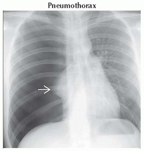
 Frontal radiograph shows typical radiographic features of tension pneumothorax. Note the hyperlucent right hemithorax and collapsed right lung
 , as well as mediastinum shifted to the left. , as well as mediastinum shifted to the left.Stay updated, free articles. Join our Telegram channel
Full access? Get Clinical Tree
 Get Clinical Tree app for offline access

|