Ultrasonography in the Evaluation of Abdominal Aortic Aneurysms


Ultrasonography in the Evaluation of Abdominal Aortic Aneurysms


Melina Vega de Céniga and Frank. A. Lederle


Medical ultrasonography was developed from principles of sonar exploration, pioneered in World War I. Sonographic images of abdominal disease were first published in 1958. This technology was widely adopted in radiology, cardiology, and obstetrics, and by 1966 reports of detection and measurement of an abdominal aortic aneurysm (AAA) by ultrasound began to appear. In 1983, Cabellon and colleagues used ultrasonography and abdominal palpation to find AAAs in asymptomatic patients with vascular disease. In the 1990s machines were developed that were more compact and affordable and that had good image quality, and clinical point-of-care ultrasonography began to progress.


Ultrasonography uses a quartz or composite piezoelectric material that generates a sound wave when an electric current is applied. The sound wave travels through tissue and is reflected, returning to the material, which in turn receives the sound and generates a current. Early ultrasonography used a single crystal to create a one-dimensional image (A-mode). Current ultrasonography uses an array of 128 or more crystals across the face of the transducer, which generate a standard two-dimensional or gray-scale screen image (B-mode). Each crystal produces a scan line that is used to create an image or frame, which is refreshed many times per second to produce the moving image on the screen. Additional modes, including three-dimensional, four-dimensional, Doppler, and tissue Doppler modes, are now also commonly available.


The normal diameter of the infrarenal aorta varies according to gender and increases with age. In young patients without vascular disease, it measures approximately 2.0 cm in men and 1.8 cm in women. When an infrarenal aorta measures 2.5 to 2.9 cm it is considered ectatic, and an AAA is diagnosed when the infrarenal aortic diameter is 3 cm or greater or when it is 1.5 times the diameter of the suprarenal aorta.


The prevalence of AAAs is 4% to 8% for men and 0.5% to 1.5% for women older than 65 years. Only 2% to 7% of AAAs are juxtarenal or suprarenal; 97% are infrarenal. The majority of AAAs are fusiform (spindle-shaped and tapering from the middle toward each end) and are located below the renal arteries, and they can involve one or both of the iliac arteries. Other possible shapes are saccular (baglike structure protruding asymmetrically from the aorta) or cylindrical. The aneurysmal sac is commonly lined by atherosclerotic changes and/or mural thrombus.


To ultrasonographically evaluate an aortoiliac segment, the patient is scanned in the supine position after 8 to 10 hours’ fasting, with a low-frequency transducer (2.4, 3.5, or 4 MHz). Gentle pressure is applied to try to move bowel loops or gas out of the way. The aorta should be imaged as completely as possible, from the proximal segment, where the celiac trunk originates, to the distal bifurcation, and the study should include the assessment of the iliac arteries whenever possible. The examination of an AAA should be focused on determining its size, shape, location (infrarenal or suprarenal), and distance from other arterial segments. The aorta is imaged in sagittal (or paramedian longitudinal) and transverse (or axial) planes at its proximal, mid, and distal portions with B-mode and color duplex ultrasound (CDU). Sometimes the coronal plane (or frontal) can be studied too.


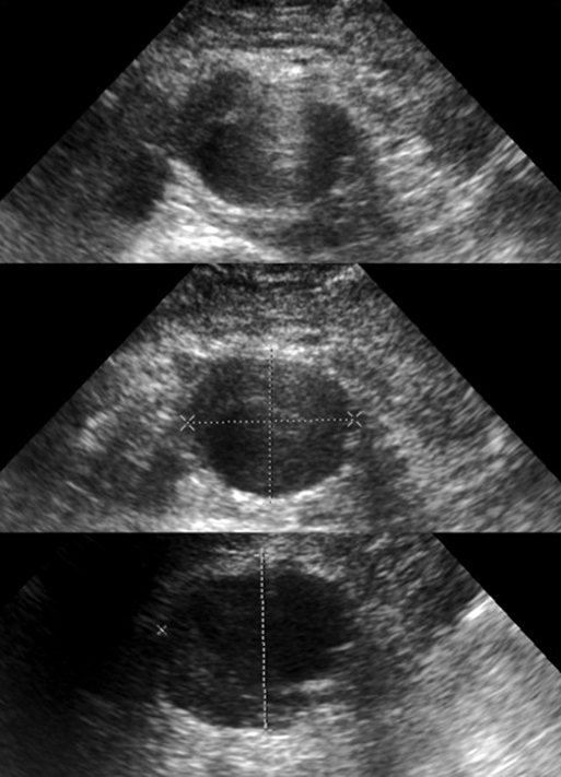
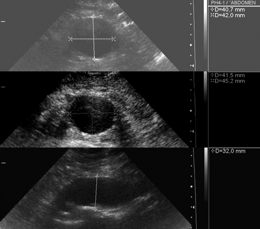
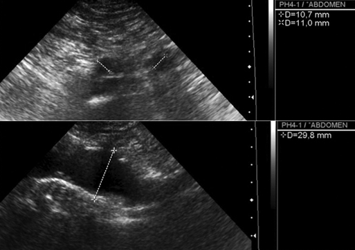
The maximum anteroposterior, transverse, and lateral aortic diameter measurements must be made from outside wall to outside wall perpendicular to the aortic axis, especially in tortuous aortas (Figure 1). Oblique measurements can overestimate the diameter. The origins of the celiac trunk, the superior mesenteric artery, and the renal arteries can all be individually documented. Careful evaluation of the course of the renal arteries and aorta in multiple planes can clarify the relation of the aneurysm to the renal arteries in case of juxtarenal AAA, although computed tomography (CT) scanning or aortography usually is required to evaluate this relationship precisely. Also, the transverse diameter of the remaining perfused lumen and the thickness and distribution (circumferential or eccentric) of intraluminal thrombus can be visualized and measured (Figure 2). Flow within the AAA may be laminar or turbulent. Turbulent flow is associated with the development of mural thrombus. The ultrasonographic appearance of a ruptured AAA is that of a large, usually hypoechoic retroperitoneal fluid collection surrounding the dilated aorta. Finally, the common iliac arteries can be imaged and measured in anteroposterior, transverse, and lateral planes (Figure 3).


Unlike the thoracic aorta, for which sound reflections from the ribs and air-filled lungs interfere with imaging, the abdominal aorta is well suited to ultrasonic evaluation. Nevertheless, it can be made difficult by overlying bowel gas and obesity, especially in the region of the proximal neck of the AAA. A left lateral decubitus or oblique approach can be helpful in these patients. Visualizing the mid and lower abdominal aorta may be best by the coronal plane through either flank. Challenges include ensuring that the inferior vena cava or another fluid-filled structure not be mistaken for the aorta and ensuring that the entire diameter of the AAA is measured.


Ultrasonography is a valid and useful tool in the diagnosis of an AAA, and the assessment of its maximum diameter and length is usually quite reliable when compared to CT or magnetic resonance imaging (MRI) or intraoperative measurement. However, it often underestimates the maximum AAA diameter by more than 2 mm, a difference that can reach 5 mm in up to a third of patients. It also often overestimates proximal neck diameter and common iliac artery size, so it cannot, by itself, adequately help plan an open or endovascular repair.


Screening for Abdominal Aortic Aneurysm


AAAs fulfill the World Health Organization (WHO) criteria for implementing a worthwhile screening program (Box 1). AAAs are common in men older than 65 years. They generally develop slowly and remain asymptomatic for years until reaching a stage involving danger of rupturing. An AAA can be diagnosed by ultrasonography inexpensively, noninvasively, and with high reliability, sensitivity, and specificity, whereas physical examination alone is often inaccurate. Ultrasound is currently the preferred method for AAA diagnosis. It is noninvasive, without known risks, readily available, straightforward to learn as a focused examination, accurate, and inexpensive. The sensitivity of ultrasound scanning for AAA is 95%, and the specificity approaches 100%. Portable ultrasound can further decrease the cost of screening up to 80% and is easier to use in outpatient and primary care settings, with a reported sensitivity of 93% and specificity of 97% for diagnosis of an AAA.


Aug 25, 2016 | Posted by in CARDIOLOGY | Comments Off on Ultrasonography in the Evaluation of Abdominal Aortic Aneurysms

Full access? Get Clinical Tree

Get Clinical Tree app for offline access