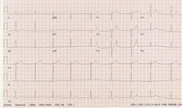
The electrocardiogram of a 57-year-old man ( Figure 1 ) was read by the computer and the over-reading cardiologist as “sinus bradycardia, otherwise normal.” The T wave in lead V 1 , however, is taller than the T wave in lead V 6 , a finding usually indicating cardiac disease. In this patient, the tall T waves in leads V 1 to V 3 likely signify posterior ischemia.

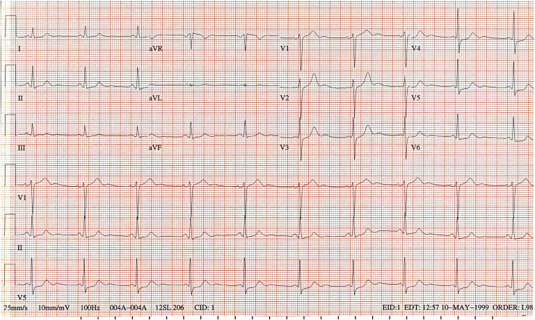
The patient had come to the hospital 12 hours earlier with new-onset angina and an electrocardiogram more clearly indicative of myocardial ischemia with ST-segment depression in leads I, II, III, aVF, and V 3 to V 6 in addition to sinus bradycardia and TV 1 taller than TV 6 ( Figure 2 ). Coronary arteriography revealed subtotal occlusion of a co-dominant left circumflex, significant narrowing of the right, and insignificant narrowing of the left anterior descending. The left circumflex and right were successfully stented.


