Tubular Mass
Jonathan H. Chung, MD
DIFFERENTIAL DIAGNOSIS
Common
Bronchiectasis (with Mucous Plugging)
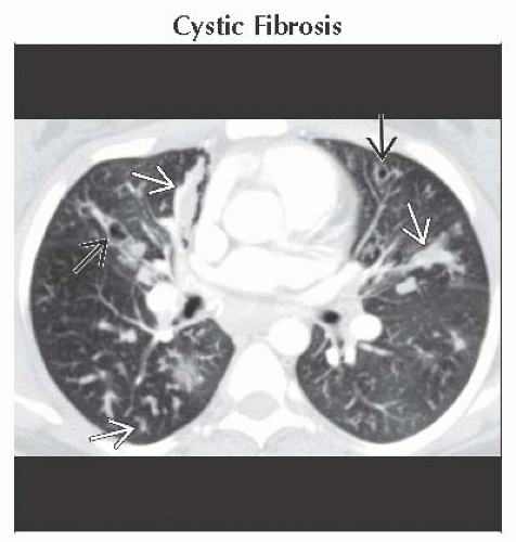
Cystic Fibrosis
Allergic Bronchopulmonary Aspergillosis
Endobronchial Tumor (with Distal Mucous Plugging)
Pulmonary Laceration
Less Common
Pulmonary AVM
Rare but Important
Scimitar Vein
Bronchial Atresia
ESSENTIAL INFORMATION
Helpful Clues for Common Diagnoses
Cystic Fibrosis
Diffuse, upper lung preponderant bronchiectasis, bronchial wall thickening
Mucous plugging in medium and large airways
Mosaic lung attenuation
Allergic Bronchopulmonary Aspergillosis
Occurs in cystic fibrosis and asthma
Central bronchiectasis in multiple lobes
Mucous-filled bronchi; may have gas-fluid level
Endobronchial Tumor (with Distal Mucous Plugging)
Slow-growing tumor, aspirated foreign body, broncholithiasis
Results in distal bronchiectasis ± mucous plugging, air-trapping
Pulmonary Laceration
Tubular lacerations more common with penetrating injuries (e.g., knife, bullets)
Laceration initially blood-filled (hematoma)
May have oblong or tubular configuration
Helpful Clues for Less Common Diagnoses
Pulmonary AVM
Single or multiple nodules with feeding artery or arteries and draining vein
Lower lung and medial lungs
History of hereditary hemorrhagic telangiectasia
Helpful Clues for Rare Diagnoses
Scimitar Vein
Anomalous pulmonary vein from right lung; drains into IVC
Hypoplastic right lung; systemic arterial supply common; ± hypoplastic pulmonary artery
Bronchial anomalies common: Bilobed right lung, bronchial diverticula, horseshoe lung
Bronchial Atresia
Congenital atresia of segmental bronchus
Left upper lobe (most common), right upper lobe, lower lobes
Bronchocele: Mucoid impaction in obstructed bronchus
Hyperlucency and paucity of vessels within affected segment
Image Gallery
 Axial CECT shows tubular mucous plugging
 and bronchiectasis and bronchiectasis  in the lungs consistent with cystic fibrosis. in the lungs consistent with cystic fibrosis.Stay updated, free articles. Join our Telegram channel
Full access? Get Clinical Tree
 Get Clinical Tree app for offline access

|