Treatment of Renal Artery Aneurysms


Treatment of Renal Artery Aneurysms


James C. Stanley and Peter K. Henke


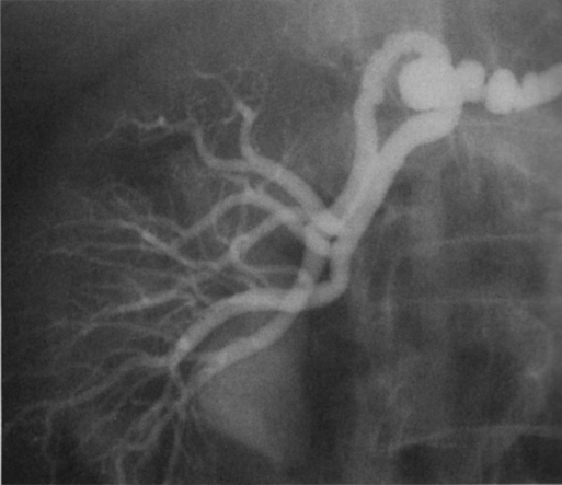
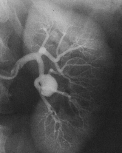
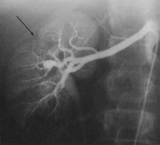
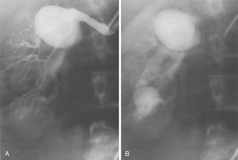
Renal artery aneurysms represent an unusual vascular disease that has been encountered with increasing regularity in clinical practice. In part, this reflects their frequent incidental recognition as a consequence of the proliferation of noninvasive imaging for nonvascular diseases. The incidence of true renal artery aneurysms approaches 0.1%, a figure derived from the 0.09% incidence of these lesions in approximately 8500 patients subjected to arteriographic studies for nonrenal disease. Women are slightly more likely than men to have renal artery aneurysms. Most renal artery aneurysms are saccular (Figure 1). Seventy-five percent are located at first- or second-order renal artery bifurcations. Intraparenchymal aneurysms occur in less than 10% of cases.


Pathogenesis


Two distinct histologic categories of aneurysms are recognized. The first is related to congenital defects, and the second is associated with arteriosclerosis. Arteriosclerotic changes are considered a secondary event rather than a primary cause of most aneurysms, but they likely cause a further loss of the vessel wall’s integrity. Most renal artery aneurysms are a consequence of preexisting congenital defects with internal elastic lamina defects or deficiencies of medial smooth muscle that can exist at bifurcations, causing the vessel wall to become functionally inadequate at withstanding normal arterial pressure with development of saccular macroaneurysms at these sites. Arterial fibrodysplasia can contribute to other aneurysms (Figure 2). Elevated blood pressures, which occur in nearly 80% of patients with renal artery aneurysms, certainly enhance the evolution of these aneurysms, but the hypertension is actually an uncommon consequence of the aneurysm itself.


Clinical Presentation


Most renal artery aneurysms are asymptomatic. An ill-defined relationship exists between renal artery aneurysms and elevated arterial blood pressure. In an occasional patient, an aneurysmal thrombus embolizes or propagates and occludes a distal artery, thereby producing renal ischemia and renovascular hypertension (Figure 3). Computational modeling suggests that deformation of branches adjacent to aneurysms at bifurcations can produce a pressure gradient and also cause renovascular hypertension. It has also been hypothesized that energy dissipation in large aneurysms can cause a similar drop in renal blood pressure and result in systematic blood pressure increases as a result of renin release. No direct evidence of this has been documented in the literature. Intrinsic stenotic disease adjacent to an aneurysm, not always evident on preoperative arteriograms, is a more likely cause of secondary hypertension in other patients, especially those with renal arterial fibrodysplasia.


Rupture occurs in less than 3% of true renal artery aneurysms. It has been suggested that aneurysms less than 1.5 cm in diameter, calcified aneurysms, and those occurring in normotensive patients are not likely to rupture, but this has not proved to be the case. Rupture may be overt into the retroperitoneal and abdominal cavity or covert into an adjacent renal vein (Figure 4).


Overt rupture causes flank pain and often hemorrhagic hypotension. It carries a reported mortality of approximately 10%. Loss of the kidney is greater than 90% following aneurysm rupture. Overt renal artery aneurysm rupture during pregnancy does not appear related to the patient’s age, presence of hypertension, or parity, and is more life-threatening. Rupture during pregnancy is associated with nearly 75% fetal mortality and 50% maternal mortality.


Treatment


Indications


Symptomatic aneurysms and those coexisting with functionally important renal artery stenoses warrant operative treatment. Asymptomatic aneurysms 2 cm in diameter, and those 1.5 to 2 cm in diameter in hypertensive patients, justify treatment by experienced interventionists. Aneurysms 1.0 to 1.5 cm in diameter provide a relative indication for elective treatment, but only when a high degree of suspicion exists that they are the cause of refractory renovascular hypertension. Because of the potential for catastrophic rupture during pregnancy, therapy is recommended for all aneurysms in women of childbearing age who might conceive in the future.


Surgical Exposure


The renal arteries are approached through an anterior abdominal, supraumbilical transverse incision. The incision is carried across both rectus muscles from the contralateral anterior axillary line to the ipsilateral posterior axillary line. A rolled sheet under the ipsilateral flank enhances operative exposure. When bilateral renal reconstructive procedures are contemplated, the same incision, extended into both flanks, is used. Transverse abdominal incisions facilitate handling of instruments in a direction perpendicular to the longitudinal axis of the body and are of particular benefit in renal artery reconstructive procedures. This technical advantage has caused transverse incisions to be favored over midline vertical incisions, although the latter incision is preferred by many surgeons.


The right renal artery and vein, as well as the inferior vena cava and aorta, are exposed by medial reflection of the colon and duodenum to the left. This exposure is accomplished by incising the lateral parietes from the hepatic flexure to the cecum and separating the mesocolon from retroperitoneal structures, usually by blunt finger dissection. The duodenum and the head of the pancreas overlying the right kidney are carefully displaced to the left as the dissection progresses. This method provides excellent visualization of the aorta, vena cava, and vessels to the right kidney. Before the renal artery dissection is begun the renal vein from its caval junction to the kidney is dissected from surrounding tissues with ligation and transection of its adrenal and ureteric tributaries. The vein may then be easily retracted during dissection of the underlying distal renal artery.


Although an aneurysm is often palpable in the hilus of the kidney, it is unwise to approach it directly. If one dissects the more-proximal renal artery first, troublesome injury to small arterial and venous branches will be lessened. Renal artery branches are usually encircled with elastic vessel loops for retraction. Vessel occlusion is best achieved with precision microvascular devices, such as Heifitz clamps.


Renal artery aneurysms occasionally are approached from behind by mobilizing the kidney and rotating it medially to expose the vessels posteriorly. In managing aneurysms involving the proximal right renal artery, exposure may be obtained by careful circumferential dissection of the inferior vena cava just below the renal veins. Entering lumbar venous branches are best ligated and transected. Retraction of the inferior vena cava then provides exposure of the renal artery at its aortic origin.


Exposure of the left renal artery follows a retroperitoneal dissection similar to that performed on the right, with reflection of the viscera, including the left colon, medially. The tail and body of the pancreas are elevated without undue tension, above the superior pole of the kidney. Only rarely does a low-lying or large spleen obscure the operative field. This retroperitoneal approach ensures much better visualization of the renal vessels than does direct exposure through an incision in the mesocolon at the root of the mesentery. Exposure of the proximal and middle portions of the left renal artery requires extensive mobilization of the overlying renal vein.

Stay updated, free articles. Join our Telegram channel

Aug 25, 2016 | Posted by in CARDIOLOGY | Comments Off on Treatment of Renal Artery Aneurysms

Full access? Get Clinical Tree

Get Clinical Tree app for offline access