Treatment of Endovascular Leaks after Aortic Endografting


Treatment of Endovascular Leaks After Aortic Endografting


Rabih A. Chaer and Michel S. Makaroun


Endovascular abdominal aortic aneurysm repair (EVAR) is predicated on complete exclusion of the aneurysm from the arterial circulation. An endoleak, or continued arterial perfusion of the aneurysm sac after endovascular treatment, can compromise the effectiveness of EVAR.


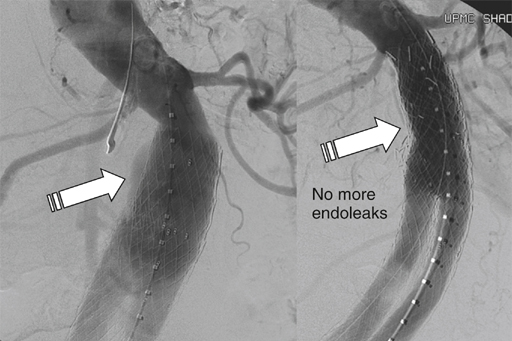
There are five major categories of endoleaks (Table 1). Some appear to be an unavoidable consequence of placing a stent graft inside an aneurysm sac with patent outflow vessels, some are the result of poor seal at the proximal or distal fixation sites or between the graft components, and others occur as a result of graft material failure. Endoleaks are associated with a variable increase in intrasac pressure that depends on the type of endoleak, which determines its severity and clinical significance.


Type I Endoleak


Type I endoleak continues to be a major cause of rupture after EVAR and should be considered a failure of the treatment. They are best prevented by proper patient and device selection. The incidence of type I endoleak increases with difficult anatomic situations, typically when devices are used outside the manufacturer’s instructions for use, such as short or angulated necks and landing zones with heavy calcifications. Type I endoleak is associated with significant pressure elevation in the aneurysm sac and has been linked to a continued risk of rupture, with a high of 22% mortality from rupture reported in earlier series. The risk of rupture from an untreated type I endoleak has been reported to be 3.4% at 15 months in the EUROSTAR (EUROpean Collaboration on Stent/graft Techniques for Aortic aneurysm Repair) registry, similar to the risk of rupture of an untreated abdominal aortic aneurysm (AAA). Whenever feasible, all type I endoleaks should be corrected promptly because spontaneous resolution, though possible, is not typical, and treatment can be simple with endovascular means. The initial treatment of procedural type I endoleak typically involves reballooning of the fixation sites, although additional aortic cuffs or iliac limb extensions, or placement of a balloon-expandable Palmaz stent (Cordis, Johnson & Johnson, Warren, NJ) are sometimes required.


Late type I endoleaks can occur as a result of graft migration, aneurysmal degeneration of the aortic neck, enlargement of the iliac arteries, or severe angulation at the fixation site that can disengage the stent graft from the aortic wall as the sac starts to shrink. The treatment of late type I endoleaks can be more challenging, but it usually also involves ballooning, stent graft extension (Figure 1), or use of Palmaz stent to increase the graft radial force and apposition to the aortic wall.


In situations where visceral branches preclude graft extension, glue or coil deposition in the track between the stent graft and the aortic wall may be successful. At the proximal attachment site, the edge of the graft can be probed with a reverse or simple-curve catheter, depending on the approach. Unless the leak is a result of gross undersizing or severe neck angulation, it often consists of a small channel alongside the graft leading to the perigraft space. A microcatheter can be advanced into this channel and embolization can be performed with microcoils, glue, or other embolic agents. It is essential to initially perform angiography of the aneurysm sac to rule out a combined type I and type II endoleak, which is not uncommon. In the case of a combined endoleak, the feeding vessel should be embolized with microcoils before the attachment site leak is treated, in order to optimize the seal and prevent adverse events from end organ damage from embolization to the colon or spinal cord. Embolization is less often required for distal type I endoleaks because a distal seal can be achieved with graft extension, and a variety of surgical or endovascular maneuvers are available to preserve hypogastric perfusion.


Open conversion is almost never required at the time of the initial EVAR, but it is sometimes required for delayed endoleak. It can be associated with increased morbidity and mortality, although several series report results that are comparable to de novo open AAA repair. This can be performed by way of a transperitoneal or retroperitoneal approach, with the initial proximal aortic control often at the supraceliac or suprarenal location, depending on the graft location and the presence of suprarenal fixation. Stepwise distal clamping is performed to reduce ischemic time, with complete or partial endograft removal. Partial explantation with in situ replacement can be performed in the absence of graft infection, with the proximal suture line incorporating the suprarenal stents, and the distal suture line of a tube or an aortoiliac graft incorporating the iliac limbs.


In one series reporting on the outcomes of reinterventions after EVAR, 51 delayed type I endoleaks with and without stent graft migration were treated by proximal stent graft extensions in 19 patients, translumbar coil embolization in 20 patients, or open surgical conversion with stent graft explants and aortoiliac reconstruction in five patients. Placement of a balloon-expandable Palmaz stent at the aortic neck was required in 14 patients, and type I endoleaks in eight patients sealed spontaneously and did not require any further intervention. Two postoperative deaths (3.9%) were reported, one from an aortoenteric fistula that developed as a procedural complication after translumbar coil embolization, and one after graft explant with aortoiliac reconstruction in a patient with prior stent graft and a Palmaz stent. Most patients can therefore be managed by endovascular means, and open conversion is sometimes required with acceptable overall morbidity and mortality.

Stay updated, free articles. Join our Telegram channel

Aug 25, 2016 | Posted by in CARDIOLOGY | Comments Off on Treatment of Endovascular Leaks after Aortic Endografting

Full access? Get Clinical Tree

Get Clinical Tree app for offline access