Treatment of Arterial Complications in Thoracic Outlet Syndrome


Treatment of Arterial Complications in Thoracic Outlet Syndrome


Enrique Criado


Arterial thoracic outlet syndrome (TOS) can be defined as the anatomic condition that produces pathologic compression of the subclavian or axillary arteries in their course through the thoracic outlet. Arterial compression can occur as an anatomic state in which the artery is externally compressed in neutral position, which with the repetitive motion of the upper extremity accentuates the mechanical stress on the arterial wall, causing its eventual injury. In other cases the artery may not be compressed in neutral position, but the repetitive arm motion suffices to cause the arterial injury. This latter mechanism is likely the culprit of arterial pathology in persons with vigorous upper extremity use, such as athletes or some laborers.


Positional or fixed arterial stenosis at the thoracic outlet, with or without poststenotic arterial dilatation, is the precursor of aneurysm formation. The development of arterial dilation is secondary to arterial wall vibration from turbulent flow dynamics, but it may be reversible following resolution of the stenosis. The natural history and clinical significance of arterial compression in patients with TOS is not well known, but in many cases it is not benign. During the early stages of arterial TOS, the artery is histologically intact, showing external compression only during arm motion, typically with arm elevation. Eventually, this dynamic compression results in post-stenotic dilatation and eventually aneurysmal degeneration of the wall. In other cases, the arterial injury results in intimal thickening and disruption that can produce fixed arterial stenosis, occlusion, or embolic phenomena.


Arterial TOS is considered the least common form of TOS and is estimated to constitute less than 5% of all TOS cases. However, the incidence of arterial TOS is probably underestimated because its diagnosis is rarely suspected unless it manifests with an ischemic complication. The University of Michigan experience with more than 400 cases of TOS revealed 17% to be the arterial type.


Anatomic Features


The subclavian artery is typically compressed as it courses between the first rib inferiorly, the anterior scalene muscle anteriorly, and the middle scalene muscle and the brachial plexus posteriorly. Arterial TOS occurs much less commonly as a result of compression of the axillary artery by the pectoralis minor muscle anteriorly or by the humeral head posteriorly. The fact is that most cases of arterial TOS are associated with the presence of cervical ribs, anomalous first ribs, or deformities secondary to clavicular or rib fractures or orthopedic surgery.


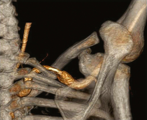
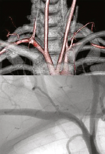
Complete cervical ribs typically elevate the floor of the thoracic outlet, significantly increasing the compression of the subclavian artery by the anterior scalene muscle (Figure 1). Cervical ribs are found in less than 0.5% of the general population, and are twice as common in women as in men. Their presence is associated with histopathologic changes in the brachial plexus, including fibrosis, vascular hyalinization, mucinous degeneration, and intraneural collagen nodules. Changes are not seen in nerve specimens of patients without cervical ribs. This observation suggests that in cases of arterial compression by a cervical rib, nerve compression is likely also present.


The incidence of cervical ribs reported in large surgical series of neurogenic TOS is between 4% and 5%. In my experience with patients operated on for all types of TOS, the overall incidence of cervical rib or other bony anomaly was 25%, whereas it was 60% among patients with arterial TOS. This difference might reflect practice referral patterns, but may be a result of more stringent surgical criteria for neurogenic TOS.


Clinical Presentation


In young patients with symptoms of acute or chronic upper extremity ischemia, the diagnosis of arterial TOS should be always entertained. The most obvious clinical presentation of TOS is acute upper extremity ischemia with a subclavian artery aneurysm. However, unless frank ischemia occurs, many patients with arterial TOS are asymptomatic. Some have subtle extremity symptoms that do not suggest arterial TOS until imaging studies support such a diagnosis. For this reason, most reports dealing with arterial TOS almost exclusively include cases presenting with arterial embolization.


In my experience with 41 patients operated on for arterial TOS, 12 patients experienced subclavian aneurysms; 24 had severe positional arterial stenosis, with poststenotic dilatation in half of the cases; two had fixed subclavian artery stenoses; two had luminal filling defects; and one had subclavian arterial occlusion. Clinically, 13 patients (32%) presented with acute ischemia, 11 of whom harbored a subclavian aneurysm. Chronic arm symptoms affected 27 patients, of whom 24 had subclavian positional stenosis and/or poststenotic dilatation, one had chronic subclavian occlusion, and two had axillary artery compression. One patient with a subclavian aneurysm had no symptoms.


Some patients complain of chronic symptoms difficult to differentiate from those of neurogenic compression. Arterial and neurogenic compression within the thoracic outlet are not always separate entities. The tight anatomic relationship between the subclavian and axillary arteries with the brachial plexus is such that in many cases both structures, although to different degrees, are pathologically compressed. For this reason, in some patients, arterial compression may be diagnosed and treated as neurogenic TOS. In the absence of acute arterial complications, significant arterial compression of the subclavian or axillary artery can produce symptomatology suggestive of upper extremity claudication, mainly with the use of the arm in elevated positions. Occasionally, patients experience hand and forearm symptoms from minor embolic events that are often misdiagnosed, unless an arterial evaluation is conducted.


The pathologic vascular changes that occur in patients with arterial TOS are progressive and can be classified in stages. Stage 1 patients have severe or complete arterial obstruction seen only with positional changes of the upper extremity, typically with arm abduction and external rotation, with the artery appearing normal in neutral position. Stage 2 patients experience arterial changes in the neutral position in the form of mild poststenotic dilatation, fixed stenosis, or luminal irregularities. Stage 3 patients have true arterial aneurysms or with complete occlusion of the subclavian or axillary arteries (Figures 2 and 3). Although it is unusual, some patients with axillary artery compression have aneurysms at the origin of the circumflex humeral arteries.


Stay updated, free articles. Join our Telegram channel

Aug 25, 2016 | Posted by in CARDIOLOGY | Comments Off on Treatment of Arterial Complications in Thoracic Outlet Syndrome

Full access? Get Clinical Tree

Get Clinical Tree app for offline access