Thyroid Goiter
Presentation
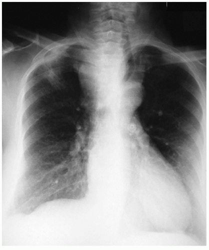
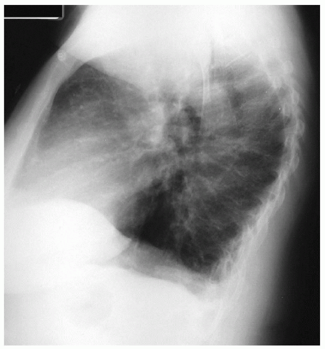
A 60-year-old woman presents to her family doctor complaining of a persistent cough for the past 3 weeks. After a short course of antibiotics, the following chest x-rays are obtained.
Case Continued
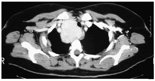
The patient’s family doctor orders a computed tomography (CT) scan of the chest and refers her to a thoracic surgeon. Upon questioning, she admits to having some difficulty swallowing solid food. The patient denies stridor, wheezing, hoarseness, weakness, and flushing. On examination, she has no respiratory distress. There is an enlarged right lobe of the thyroid gland, without any cervical adenopathy. The rest of the examination is unremarkable.
Discussion
This patient has a mass in the anterosuperior mediastinum. The mediastinum is composed of three regions: the anterosuperior, middle, and posterior compartments. Common tumors arising in the anterosuperior mediastinum include thymoma, lymphoma, germ cell tumor, and substernal thyroid.
The clinical presentation of a patient with a mediastinal tumor varies. About 50% of patients with mediastinal tumors are asymptomatic. Others may experience symptoms related to mass effect, tumor invasion, or hormonal production. Hoarseness and Horner’s syndrome are usually associated with malignancy. Superior vena cava syndrome and airway compression may be caused by large tumors.
▪ CT Scans
 Figure 7-3
Stay updated, free articles. Join our Telegram channel
Full access? Get Clinical Tree
 Get Clinical Tree app for offline access

|

