Figure 2.1
(a, b) Anterior thoracotomy. The patient is placed in the lateral decubitus position with the back toward the edge of the table, with the middle of the table flexed. The patient is rolled at a 45° angle posteriorly for best anterolateral hemithorax exposure. The skin incision follows the fourth or fifth intercostal space horizontally beginning parasternally to the midaxillary line two fingerbreadths below the tip of the scapula. In women, the incision is made in the submammary fold and along the retromammary space between the deep fascia and the breast. The pectoral and anterior serratus muscles are incised in an avascular plane, and the hemithorax is opened in the fourth intercostal space. The anterior border of the latissimus dorsi muscle is identified and incised by electrocautery to facilitate further access. Care should be taken to avoid injury to the long thoracic nerve. Segments of 1 cm in one or two cartilages may be transected and removed in patients with a rigid thoracic cage in whom rib fractures or luxation may be anticipated. The neuromuscular bundles are divided and transfixation ligated to avoid postoperative bleeding. A rib-spreading retractor is placed between the ribs, and the intercostal muscles are detached from the superior border of the fifth rib as posteriorly as possible. The horizontal incision allows enlargement posterolaterally if further access is essential to the operation. For closure of the anterolateral thoracotomy, as also is true for posterolateral thoracotomy, pericostal absorbable sutures are placed, and the intercostal muscle may be transfixed. Special diligence is needed to avoid accidental laceration of the pericardium or heart by placing the anterior pericostal suture on the left side. After careful closure of the anatomic layers of the greater pectoral, anterior serratus, and latissimus dorsi muscles, subcutaneous drains may be placed to avoid fluid collection

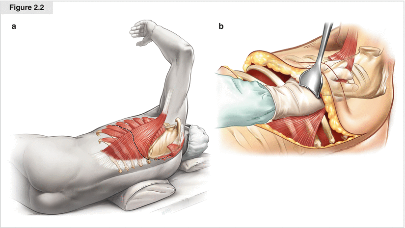
Figure 2.2


(a, b) Posterolateral thoracotomy. The patient is placed in the lateral decubitus position at a 90° angle by flexing the middle of the table or placing a sandbag under the operating table mattress. The curvilinear incision starts in front of the axillary line and goes one fingerbreadth below the inferior tip of the scapula; it then proceeds in a posterosuperior direction between the posterior border of the scapula and vertebral column. There, it may be extended up to the neck at level TH1. If further exposure of the anterolateral chest wall is needed, the skin incision may be extended anteriorly to the inframammary crease. If resection of the carina or main bronchus via a lateral thoracotomy is planned, opening the chest wall through the fourth intercostal space is recommended to achieve the best exposure of the central bronchial system. Diathermy is used to transect the fibers of the lower trapezius muscle diagonally and those of the latissimus dorsi muscle anteriorly. The posterior border of the anterior serratus muscle may be incised, or the muscle may be retracted anteriorly. The lower portion of the greater rhomboid muscle, with its medial insertion on the medial aspect of the scapula and the insertion of the anterior serratus muscle at the lower and medial edges, is divided, and a scapula retractor is placed under the serratus fascia and scapula. This allows the surgeon to pass the hands up paraspinally for numbering the ribs. At the uppermost hemithorax, attachments of the posterior serratus muscle may be palpated, which may help identify the first rib. For standard resections, the fifth intercostal space is identified and division of the intercostal muscles from the superior border of the back end opens the chest cavity. The intercostal dissection should be extended as anteriorly as possible, allowing further spreading of the ribs without fracture. Posteriorly, the paraspinous muscle is dissected down to the costovertebral junction. To avoid rib fracture by spreading the ribs, resection of a 2-cm portion of the superior or inferior rib at the costovertebral angle is recommended. Transfixation ligatures or electrocautery of the intercostal bundle is advisable, because it may be a potential source of postoperative bleeding complications. The pleural space is opened with care if adhesions are expected. Once the rib spreader is in place, it should be opened slowly to minimize the risk of rib fracture. Closure of the incision begins with blockage of the third to seventh intercostal nerves with injection of a local anesthetic if no epidural or paravertebral catheter is in place. After the operating table is re-flexed and repositioned, usually six pericostal heavy absorbable sutures are placed. The edges of the anterior serratus and greater rhomboid muscles are reattached to the scapula, and the diverted latissimus dorsi and trapezius muscles are repaired. In most patients, the extent of the incision requires subcutaneous drain placement to prevent fluid collection
Stay updated, free articles. Join our Telegram channel

Full access? Get Clinical Tree


