Figure 1.1
Standard positioning of the patient for VATS is lateral decubitus with flexion of the torso. The point of flexion should be about two fingers below the tip of the scapula. The arm should be positioned to ensure that the shoulder forms the highest point; the upper arm is placed as low as possible so that movement of the camera and instruments is as unimpeded as possible. Skin preparation with chlorhexidine- or povidone-iodine–based solutions and sterile draping are done in the same way as for an anterolateral thoracotomy. This allows for easy conversion to standard thoracotomy if needed. The surgeon is positioned either on the ventral side (e.g., to evaluate pleural effusion or empyema) or at the dorsal side (e.g., to assess a mediastinal tumor or pericardial effusion) of the patient
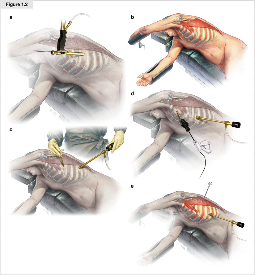
Figure 1.2


(a) Uniport thoracoscopy: the combined camera and working channel inserted through single incision. (b) The first incision is chosen based on the indication for surgery and the anatomic structures. As a rule, the muscle-free axillary triangle bordered by the axilla, the dorsal edge of the greater pectoral muscle, and the ventral edge of the latissimus dorsi muscle is an ideal area. It must be chosen so that the total pleural cavity can be explored and extensions of the incision can be done easily if necessary. (c–e) The number and placement of the other ports depends on the individual indication and ranges from one—mostly in the sixth intercostal space—to two incisions for evaluation of pleural effusion (c) to three or four for a VATS-lobectomy (e). The standard is two to three ports (pneumothorax), with positioning as in (d). The surgeon chooses a 5- or 10-mm trocar for the camera and one to two working ports for grasping forceps, minimally invasive surgery scissors, endoscopic suction, endoscopic excision forceps, endoscopic tweezers, and eventually the endoscopic stapler. The use of ports in VATS is optional except for the camera port. The chest tube (24–28F) should be placed via the most caudal incision (sixth to seventh intercostal space). The other incisions should be closed with two-layered absorbable sutures
Stay updated, free articles. Join our Telegram channel
Full access? Get Clinical Tree
