Fig. 3.1
Positioning for seated anterior–posterior esophagram. There is a disk of known diameter taped to the right side of the neck, a towel protecting the clothing from spilled barium, and a lead apron shielding the reproductive organs
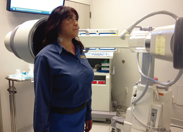
Fig. 3.2
Positioning for standing anterior–posterior esophagram
Table 3.1
Esophageal screen protocol
Anterior–posterior fluoroscopic view |
20 ml 60 % w/v liquid barium |
13 mm barium tablet |
Comprehensive VFE Technique
The patient is first evaluated upright in the AP view similar to the ES . We prefer the upright position for the initial swallows in order to evaluate the esophagus in a natural eating position with the benefit of gravity. The patient is positioned on a chair in the AP view (Fig. 3.1). The patient is given a lead shield to protect the pelvic region and obstructing clothing and jewelry are removed. A towel is draped over the shoulders and lap to prevent drips of barium on the patient’s clothing. The patient is then given 20 ml of 60 % w/v liquid barium to consume via a cup without a straw. The patient is instructed to take the largest sip comfortable from the cup and consume the barium with one swallow. Initiating a second swallow will arrest and reinitiate esophageal peristalsis, affecting the evaluation of esophageal body motility (deglutitive inhibition) . The patient is instructed to “swallow hard and swallow once.” The bolus is followed from the oral cavity until it enters the stomach. Once the barium has cleared the entire esophagus, the patient is given a cup with 10 ml of effervescent crystals (EZ-Gas II; E-Z-EM, Lake Success, NY) and 10 ml of water and asked to drink rapidly. A second 20 ml barium bolus with identical instructions is then administered. Collapsed and partially collapsed mucosal relief views are obtained. Once the barium has cleared the esophagus, the patient is given a 2 oz cup of water and asked to swallow a 13 mm barium tablet. The tablet helps to identify a site of obstruction with a lumen size less than 13 mm. The examination is recorded under real-time fluoroscopy at 30 fps until the tablet has entered the stomach. If there is significant delay in the passage of the barium bolus or tablet, the fluoroscopy is intermittently turned off to limit radiation exposure. The esophagus is evaluated every 60 s until the passage of the tablet or bolus or until 5 min has elapsed .
The patient is then placed recumbent on a table to acheive the right anterior oblique (RAO) projection. This position eliminates the benefit of gravity and helps identify esophageal dysmotility . The patient is placed chest down and turned to the left and instructed to rotate 40° so that the right anterior thorax is against the radiology table. The right arm is placed down at the side with the left arm flexed at the elbow and placed up by the head. The left knee is flexed and a bottle of liquid barium containing a straw is placed in their left hand (Fig. 3.3). The RAO position differentiates the esophagus from the spine and provides optimal orientation for the evaluation of esophageal anatomy and function. The patient is instructed to take the largest straw sip comfortable with similar instructions regarding the importance of a single swallow . The barium is followed from the oral cavity to its entry into the stomach . When the barium has cleared the esophagus, the patient is asked to gulp from the bottle with multiple swallows to maximally distend the esophagus. This sequential swallowing maneuver is essential to evaluate the esophagus for webs, rings, and hernias, particularly at the gastroesophageal junction (GEJ). Esophageal peristalsis should not be evaluated during the sequential swallow task because of deglutitive inhibition. If there is barium stasis in the transition zone of the esophagus (junction of proximal and middle third), the patient is asked to perform two dry swallows to relax the upper esophageal sphincter and evaluate for esophagopharyngeal reflux. If there is significant delay in the passage of the barium bolus, the fluoroscopy is intermittently turned off to limit radiation exposure. The esophagus is evaluated every 60 s with flash pedal-tap-only views until the passage of the bolus or until 5 min has elapsed .
Stay updated, free articles. Join our Telegram channel

Full access? Get Clinical Tree


